

Abdominal ultrasonographic findings of cats with feline infectious peritonitis: an update
- PMID: 38095890
- PMCID: PMC10811767
- DOI: 10.1177/1098612X231216000
Abdominal ultrasonographic findings of cats with feline infectious peritonitis: an update
Abstract
Objectives: The aim of this study was to describe the abdominal ultrasonographic findings in cats with confirmed or presumed feline infectious peritonitis (FIP).
Methods: This was a retrospective study performed in an academic veterinary hospital. The diagnosis of FIP was reached on review of history, signalment, clinical presentation, complete blood count, biochemistry panel, peritoneal fluid analysis, cytology and/or histopathology results from abnormal organs, and/or molecular testing (immunohistochemical or FIP coronavirus [FCoV] RT-PCR). Cats with confirmed FIP by molecular testing or with a highly suspicious diagnosis of FIP were included. Abdominal ultrasound examination findings were reviewed.
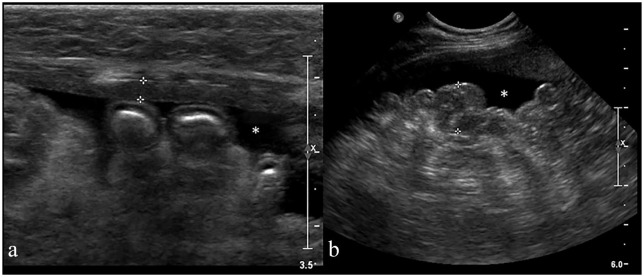
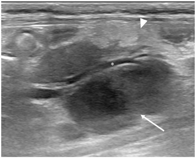
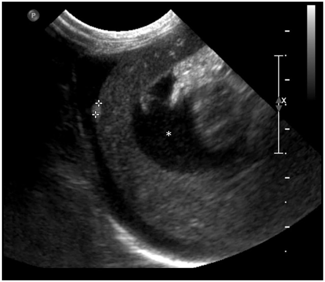
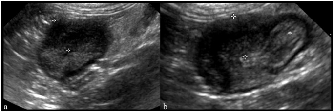
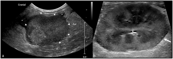
Results: In total, 25 cats were included. Common clinical signs/pathology findings included hyperglobulinemia (96%), anorexia/hyporexia (80%) and lethargy (56%). Abdominal ultrasound findings included effusion in 88% and lymphadenopathy in 80%. Hepatic changes were noted in 80%, the most common being hepatomegaly (58%) and a hypoechoic liver (48%). Intestinal changes were noted in 68% of cats, characterized by asymmetric wall thickening and/or loss of wall layering, with 52% being ileocecocolic junction and/or colonic in location. Splenic changes were present in 36% of cats, including splenomegaly, mottled parenchyma and hypoechoic nodules. Renal changes were present in 32%, encompassing a hypoechoic subcapsular rim and/or cortical nodules. Mesenteric and peritoneal abnormalities were seen in 28% and 16% of cats, respectively. Most cats (92%) had two or more locations of abdominal abnormalities on ultrasound.
Conclusions and relevance: The present study documents a wider range and distribution of ultrasonographic lesions in cats with FIP than previously reported. The presence of effusion and lymph node, hepatic and/or gastrointestinal tract changes were the most common findings, and most of the cats had a combination of two or more abdominal abnormalities.
Keywords: Ultrasound; abdomen; feline infectious peritonitis; viral diseases.
Conflict of interest statement
Conflict of interestThe authors declared no potential conflicts of interest with respect to the research, authorship, and/or publication of this article.
Figures
Similar articles
-
Sensitivity and specificity of a real-time reverse transcriptase polymerase chain reaction detecting feline coronavirus mutations in effusion and serum/plasma of cats to diagnose feline infectious peritonitis.BMC Vet Res. 2017 Aug 2;13(1):228. doi: 10.1186/s12917-017-1147-8. BMC Vet Res. 2017. PMID: 28768514 Free PMC article.
-
Diagnosis of non-effusive feline infectious peritonitis by reverse transcriptase quantitative PCR from mesenteric lymph node fine-needle aspirates.J Feline Med Surg. 2019 Oct;21(10):910-921. doi: 10.1177/1098612X18809165. Epub 2018 Nov 8. J Feline Med Surg. 2019. PMID: 30407137 Free PMC article.
-
2022 AAFP/EveryCat Feline Infectious Peritonitis Diagnosis Guidelines.J Feline Med Surg. 2022 Sep;24(9):905-933. doi: 10.1177/1098612X221118761. J Feline Med Surg. 2022. PMID: 36002137 Free PMC article. Review.
-
Abdominal ultrasonographic findings associated with feline infectious peritonitis: a retrospective review of 16 cases.J Am Anim Hosp Assoc. 2010 May-Jun;46(3):152-60. doi: 10.5326/0460152. J Am Anim Hosp Assoc. 2010. PMID: 20439937
-
Feline Infectious Peritonitis: European Advisory Board on Cat Diseases Guidelines.Viruses. 2023 Aug 31;15(9):1847. doi: 10.3390/v15091847. Viruses. 2023. PMID: 37766254 Free PMC article. Review.
Cited by
-
Thoracic radiographic findings in cats with feline infectious peritonitis.J Feline Med Surg. 2025 Feb;27(2):1098612X241309823. doi: 10.1177/1098612X241309823. J Feline Med Surg. 2025. PMID: 39930322 Free PMC article.
-
A review of feline infectious peritonitis virus infection.Vet World. 2024 Nov;17(11):2417-2432. doi: 10.14202/vetworld.2024.2417-2432. Epub 2024 Nov 5. Vet World. 2024. PMID: 39829669 Free PMC article. Review.
-
Ultrasonographic Renal Subcapsular Thickening in Cats with Primary and Metastatic Carcinoma.Vet Sci. 2024 Mar 20;11(3):134. doi: 10.3390/vetsci11030134. Vet Sci. 2024. PMID: 38535868 Free PMC article.
References
-
- Rohrbach BW, Legendre AM, Baldwin CA, et al.. Epidemiology of feline infectious peritonitis among cats examined at veterinary medical teaching hospitals. J Am Vet Med Assoc 2001; 218: 1111–1115. - PubMed
MeSH terms
LinkOut - more resources
Full Text Sources
Miscellaneous
