. 2024 Apr 9;8(7):1820-1833.
doi: 10.1182/bloodadvances.2023011766.

Genetically corrected RAG2-SCID human hematopoietic stem cells restore V(D)J-recombinase and rescue lymphoid deficiency

Affiliations

Genetically corrected RAG2-SCID human hematopoietic stem cells restore V(D)J-recombinase and rescue lymphoid deficiency

Mara Pavel-Dinu et al. Blood Adv. .

Abstract

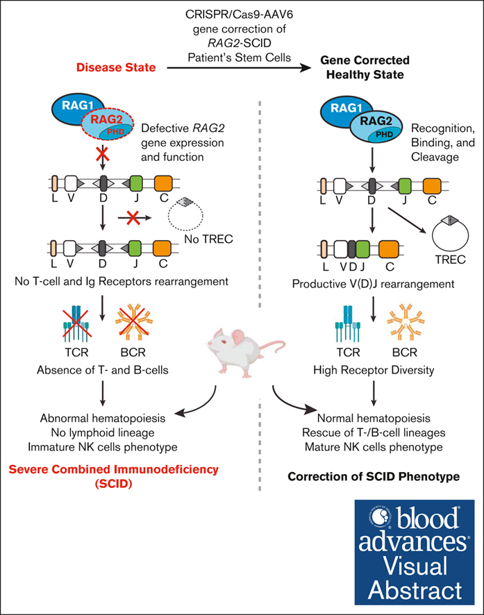
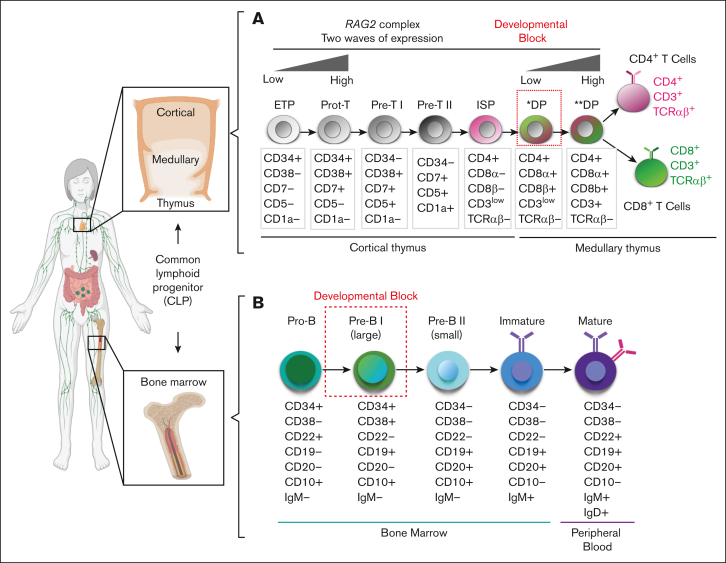
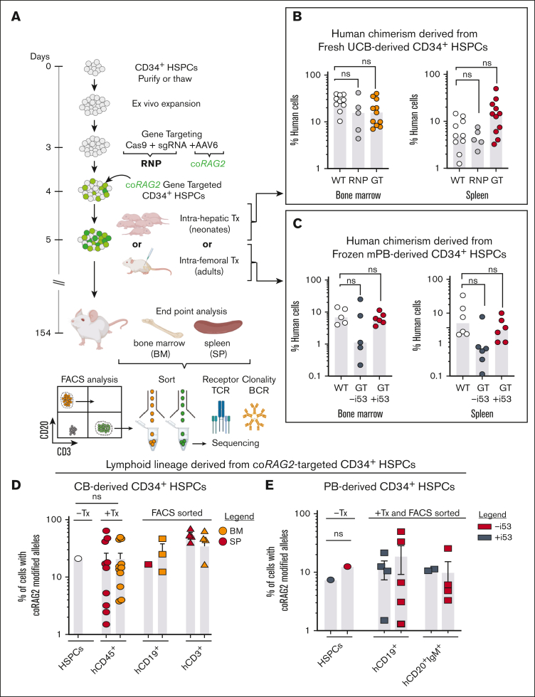
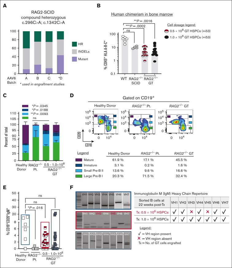
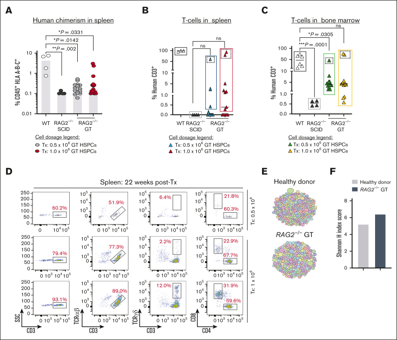
Recombination-activating genes (RAG1 and RAG2) are critical for lymphoid cell development and function by initiating the variable (V), diversity (D), and joining (J) (V(D)J)-recombination process to generate polyclonal lymphocytes with broad antigen specificity. The clinical manifestations of defective RAG1/2 genes range from immune dysregulation to severe combined immunodeficiencies (SCIDs), causing life-threatening infections and death early in life without hematopoietic cell transplantation (HCT). Despite improvements, haploidentical HCT without myeloablative conditioning carries a high risk of graft failure and incomplete immune reconstitution. The RAG complex is only expressed during the G0-G1 phase of the cell cycle in the early stages of T- and B-cell development, underscoring that a direct gene correction might capture the precise temporal expression of the endogenous gene. Here, we report a feasibility study using the CRISPR/Cas9-based "universal gene-correction" approach for the RAG2 locus in human hematopoietic stem/progenitor cells (HSPCs) from healthy donors and RAG2-SCID patient. V(D)J-recombinase activity was restored after gene correction of RAG2-SCID-derived HSPCs, resulting in the development of T-cell receptor (TCR) αβ and γδ CD3+ cells and single-positive CD4+ and CD8+ lymphocytes. TCR repertoire analysis indicated a normal distribution of CDR3 length and preserved usage of the distal TRAV genes. We confirmed the in vivo rescue of B-cell development with normal immunoglobulin M surface expression and a significant decrease in CD56bright natural killer cells. Together, we provide specificity, toxicity, and efficacy data supporting the development of a gene-correction therapy to benefit RAG2-deficient patients.


Conflict of interest statement

Conflict-of-interest disclosure: M.H.P. holds equity and is on the board of directors of Graphite Bio; is on the scientific advisory board and holds equity in Allogene Therapeutics; and is a founder and holds equity in Kamau Therapeutics. R.M. is on the board of directors of Beyond Spring Inc, and the scientific advisory boards of Zenshine Pharmaceuticals, and Kodikaz Therapeutics Solutions Inc; none of these companies had any input in the design, execution, interpretation, or publication of the work in this manuscript. G.L.K., N.M.B., and C.A.V. are employees of integrated DNA technologies, which offer reagents for sale that are identical or similar to some of the compounds described in the manuscript. The remaining authors declare no competing financial interests.

Figures

None
Graphical abstract
Figure 1.
Figure 1.
Schematics of hematopoietic developmental defect for patients with RAG2-SCID. (A) Overview of human T-cell (top) and (B) B-cell (bottom) developmental stages in the thymus and BM from cCLP. Dotted red squares mark developmental block for patients with RAG2-SCID. ∗DP, preselection double-positive stage; ∗∗DP, postselection double-positive stage; ETP, early thymic progenitor; ISP, immature single-positive stage.
Figure 2.
Figure 2.
Efficacy and specificity of genome editing at the RAG2 locus using universal correction strategy. (A) Schematics of gene-targeted integration of codon- coRAG2 and an expression cassette. The coRAG2 sequence is under the control of an endogenous promoter. (B) Overview of genome-editing outcomes. (C) Percent coRAG2-GTed in healthy donor (HD)purified HSPCs from fresh CB and frozen PB. Each circle represents a unique human HSPC donor. Genome-targeted (GTed) integration of coRAG2 was quantified by digital-droplet polymerase chain reaction (ddPCR). (D) Frequency of cells with 1 (monoallelic) or 2 (biallelic) alleles targeted as a function of the virus’ MOI. Analysis was performed on single cells sorted on methylcellulose plates (n = 5 at 5000 MOI; n = 4 at 2500 MOI; and n = 2 at 1250 MOI). Bars ± SEM (E) OT analysis using RAG2-SCID (c.296C>A and c.1342C>A) patient-derived HSPCs. Next-generation sequencing of 48 COSMID predicted OT sites in edited-only (RNP-sgRNA guide no. 3 and HiFi Cas9 nuclease) or electroporated-only (mock, nucleofected without RNP) cells. Shown INDELs reads for on-target (RAG2 gene, white circle) and OT sites below the limit of detection (gray circles) and above the limit of detection (green circles). PacBio-based analysis overview of (F) quantification of perfect and imperfect homologous recombination events using PacBio sequencing. Marked as “wild-type∗” are events that retained the wild-type protospacer adjacent motif sequence even after perfect HR repair. (G) A total of 85.5% of the bulk non-GT alleles have INDELs and 14.5% are wild-type (WT) alleles. The brown bar represents the background signal from sequencing error. (H) An overview of the INDEL spectrum in non-GT alleles identified short deletions (∼13 bp) as the predominant events. Each dot represents the frequency of a unique PacBio long-read size bin. Red box shows ≥100 bp deletions in (I) top histogram. Blue box shows ≥100 bp insertions in (I) bottom histogram. Percent total frequency of ≥100 bp deletions is 4.2 and of ≥100 bp insertions is 4.0 (supplemental Figure 4D). SNP, single nucleotide polymorphism.
Figure 3.
Figure 3.
In vivo lymphoid lineage development from coRAG2-targeted healthy donor-derived HSPCs. (A) Schematic of engraftment protocol of human HSPCs into NSG immunodeficient mice and secondary analysis. (B) Percent human cell chimerism (CD45+ HLA A-B-C+ double-positive cells) in bone marrow (BM) and spleen (SP) of mice 22 weeks posttransplant with CB (intra-hepatic [IH] transplant [Tx]) or (C) PB-derived HSPCs (intra-femoral [IF] transplant [Tx]) edited with RAG2-sgRNA guide 3 (RNP, light grey circles in panel B) or targeted with coRAG2 cassette (GTed, yellow and red circles in panel B, dark grey and red circles in panel C). Each dot represents an individual mouse; panel B: WT (n = 10), RNP (n = 5), GTed (n = 11); panel C: WT (n = 5), GTed-i53 (n = 5), GTed+i53 (n = 6). Bars, median (D) percent coRAG2-GTed by ddPCR before (-Tx) and after (+Tx) transplant of CB and (E) PB-derived CD34+ HSPCs into NSG mice and FACS sort from BM, as marked. Bars, mean ± s.e.m.; -i53, without p53 inhibitor; +i53, with p53 inhibitor; stats. One-way ANOVA, nonparametric test, Kruskal-Wallis test, Dunn’s multiple comparisons test.
Figure 4.
Figure 4.
Ex vivo gene-targeted RAG2null HSPCs correct in vivo B cell developmental block. (A) Percent of total genome editing (INDELs and HR) in RAG2null patient-derived HSPCs, using 4 different AAV6 production lots. AAV6 lot D (asterisk marked) was used for all subsequent engraftment studies. (B) Percent human cells (CD45+ HLA A-B-C+) engrafted in BM after transplanting (22 weeks post-Tx) 0.5 x10⁶ uncorrected RAG2null patient-derived HSPCs (n=4 mice, grey circles), 0.5 x10⁶ coRAG2-GT HSPCs (n=15 mice, half red circles) or 1.0 x10⁶ coRAG2-GT HSPCs (n=15 mice, black circles). Healthy donor (HD) HSPCs were used as control (n=6, white circles). RAG2null patient genotype: c.296C>A; c.1342C>A. (C) FACS-based quantification is shown in (D) of large Pre-B I, small Pre-B II, immature, and mature B cells derived from coRAG2-GT HSPCs. Each population is graphed as a percent of total B cells. Bars: mean ± s.e.m. (D) Representative FACS plots of B-cell developmental stages derived from a healthy donor (left panel), RAG2null patient (middle panel), and coRAG2-GT RAG2null HSPCs. FACS-based quantification of percent cells in each developmental stage is shown. (E) FACS-based quantification of CD19+CD20+IgM+ triple-positive B cells derived from each condition tested. (F) PCR-based sequencing of immunoglobulin M (IgM) heavy chain (Vh) families from sorted triple-positive B-cells.
Figure 5.
Figure 5.
Correction of RAG2 gene function in RAG2null HSPCs restores V(D)J activity and normal T cells development. (A) Percent human cells (CD45+ HLA A-B-C+) detected in spleen (SP) (22 weeks post-Tx) with coRAG2-GTed RAG2null HSPC (0.5 x 10⁶, grey circles or 1.0 x 10⁶, red circles). HD (white circles) and uncorrected RAG2-/- (black circles) HSPCs-derived human cells were engrafted and analyzed in parallel. (B) Human CD3+ T-cells detected in the spleen (SP) and (C) bone marrow (BM) derived from coRAG2-GTed RAG2null HSPCs. (D) FACS plots showing T-cells analysis derived from 3 mice with the highest level of human CD3+ cells (dotted squares). Functional V(D)J rearrangement is demonstrated by the presence of CD3+TCR α/β, CD3+TCR γ/δ, and single-positive CD4+ and CD8+ derived from coRAG2-GTed RAG2null HSPCs. (E) Treemap diversity analysis for TCRA/TCRD CDR3 sequences from sorted CD3+ cells from (C). Each circle is a unique CDR3 sequence, and the size of the circle represents the frequency out of the total number of reads. (F) Shannon H index score quantification of CDR3 sequence from (E-F), showing oligoclonal repertoire. Shannon index score of ≥8 indicates polyclonal repertoire. Stats. One-way ANOVA, nonparametric, Kruskal-Wallis test. Median plotted in A-C.

Comment in

References

    1. Fischer A, Notarangelo LD, Neven B, Cavazzana M, Puck JM. Severe combined immunodeficiencies and related disorders. Nat Rev Dis Primers. 2015;1 - PubMed
    1. Sobacchi C, Marrella V, Rucci F, Vezzoni P, Villa A. RAG-dependent primary immunodeficiencies. Hum Mutat. 2006;27(12):1174–1184. - PubMed
    1. Notarangelo LD, Kim MS, Walter JE, Lee YN. Human RAG mutations: biochemistry and clinical implications. Nat Rev Immunol. 2016;16(4):234–246. - PMC - PubMed
    1. Delmonte OM, Schuetz C, Notarangelo LD. RAG Deficiency: two genes, many diseases. J Clin Immunol. 2018;38(6):646–655. - PMC - PubMed
    1. Tirosh I, Yamazaki Y, Frugoni F, et al. Recombination activity of human recombination-activating gene 2 (RAG2) mutations and correlation with clinical phenotype. J Allergy Clin Immunol. 2019;143(2):726–735. - PMC - PubMed

Publication types

MeSH terms