Review
. 2023 Sep;30(5):397-406.
doi: 10.1053/j.akdh.2023.04.004.

Genetic Spectrum of Polycystic Kidney and Liver Diseases and the Resulting Phenotypes

Affiliations
Review

Genetic Spectrum of Polycystic Kidney and Liver Diseases and the Resulting Phenotypes

Hana Yang et al. Adv Kidney Dis Health. 2023 Sep.

Abstract

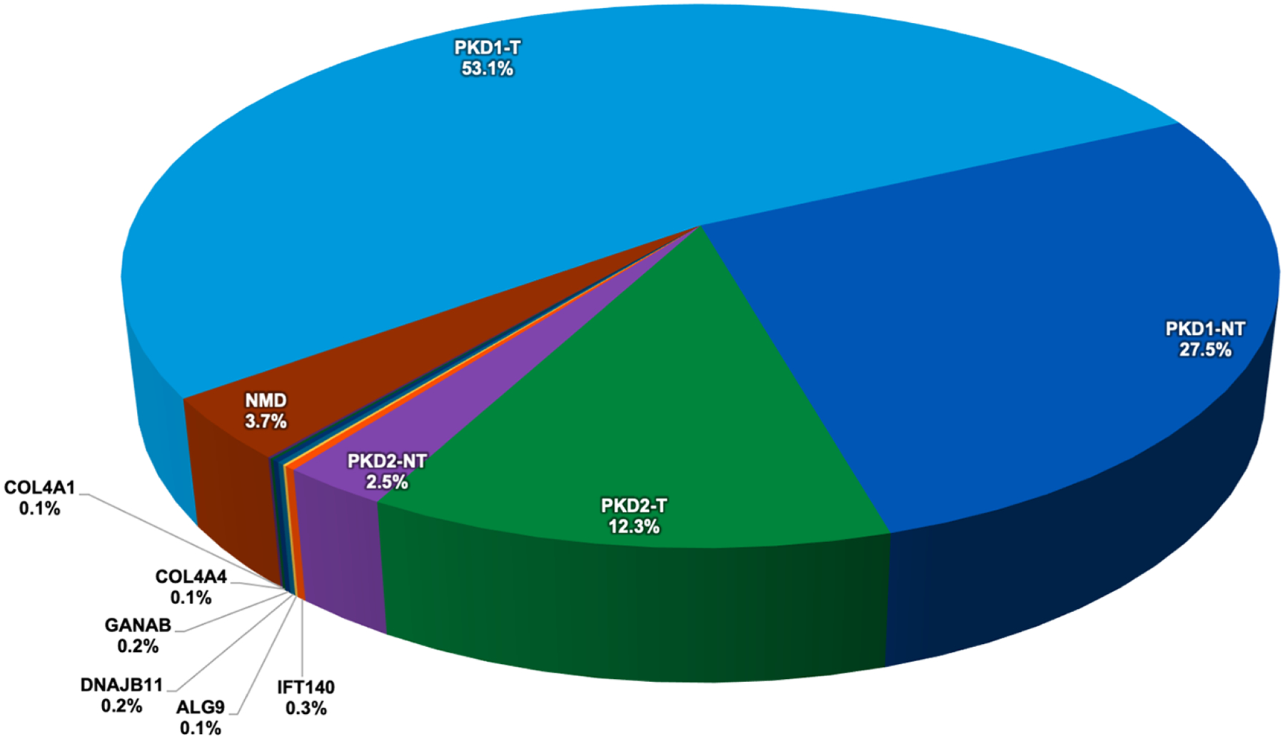
Polycystic kidney diseases are a group of monogenically inherited disorders characterized by cyst development in the kidney with defects in primary cilia function central to pathogenesis. Autosomal dominant polycystic kidney disease (ADPKD) has progressive cystogenesis and accounts for 5-10% of kidney failure (KF) patients. There are two major ADPKD genes, PKD1 and PKD2, and seven minor loci. PKD1 accounts for ∼80% of patients and is associated with the most severe disease (KF is typically at 55-65 years); PKD2 accounts for ∼15% of families, with KF typically in the mid-70s. The minor genes are generally associated with milder kidney disease, but for DNAJB11 and ALG5, the age at KF is similar to PKD2. PKD1 and PKD2 have a high level of allelic heterogeneity, with no single pathogenic variant accounting for >2% of patients. Additional genetic complexity includes biallelic disease, sometimes causing very early-onset ADPKD, and mosaicism. Autosomal dominant polycystic liver disease is characterized by severe PLD but limited PKD. The two major genes are PRKCSH and SEC63, while GANAB, ALG8, and PKHD1 can present as ADPKD or autosomal dominant polycystic liver disease. Autosomal recessive polycystic kidney disease typically has an infantile onset, with PKHD1 being the major locus and DZIP1L and CYS1 being minor genes. In addition, there are a range of mainly recessive syndromic ciliopathies with PKD as part of the phenotype. Because of the phenotypic and genic overlap between the diseases, employing a next-generation sequencing panel containing all known PKD and ciliopathy genes is recommended for clinical testing.

Keywords: ADPKD; ADPLD; ARPKD; Genetics; Polycystic kidney disease.


Conflict of interest statement

P. Harris reports the following:

Consultancy: Vertex, Mitobridge, Regulus, Otsuka, Janssen, Maze Therapeutics, CorrectorBio

Figures

Figure 1:
Figure 1:. Summary of published results from research genetic screening of the CRISP and HALT PKD ADPKD populations.
,,,,,, PKD1 truncating (PKD1-T) and PKD1 nontruncating (PKD1-NT) plus PKD2-T and PKD2-NT are the major variant types detected (95.4%), but a few individuals remain unresolved (no mutation detected; NMD), and minor genes account for ~1% of cases.
Figure 2:
Figure 2:. Summary of the types of pathogenic variants described from genetic screening of the PKD1 and PKD2 genes and collated from the ADPKD Variant Database (pkdb.mayo.edu).
(A) Truncating mutations cause a shorter form of the protein by shifting or blocking the reading frame while nontruncating mutations usually produce a full-length but altered protein. Canonical and noncanonical splicing mutations introduce disruptions to the splicing process, although the exact changes are difficult to predict. Canonical splicing involves alterations of the 5’ or 3’ dinucleotides flanking the exon (−1 G>A substitution, illustrated), while noncanonical changes are elsewhere in the intron or exon; most often involving the 5’ intronic nucleotides −12 to −3 or 3’ nucleotides +3 to +6 (+6 T>C substitution, illustrated). Frameshifting of the open reading frame is caused by base pair deletions, duplication, or insertions (duplication of TG dinucleotide, illustrated). Nonsense mutation leads to the introduction of stop codon and the early termination of the protein (C>T substitution resulting in the TAG stop codon, illustrated). CNV can be deletions, duplications, or insertions of large portions (at least one exon) of the gene (deletion of one exon, illustrated). Inframe deletions, duplications, or insertions involve changes of nucleotide number divisible by 3 that eliminate or add one or a few amino acids (TTC deletion resulting in loss of a phenylalanine amino acid from the protein, illustrated). Missense mutations substitute a single nucleotide and result in an altered amino acid in the protein (C>T change resulting in leucine to serine substitution, illustrated). (B) For PKD1 (1874 families; 81.2%), truncating variant types (frameshifting deletions/duplications/insertions [Indel], nonsense, canonical splicing, or copy number variant [CNV]) account for 51.3% of the total, while nontruncating (inframe deletions/duplications/insertions [Indel], missense, and noncanonical splicing) represent 29.9%. For PKD2 (434 families; 18.8%), truncating variants account for 16.0% and nontruncating 2.8% of the total.
Figure 3:
Figure 3:. General scheme for identifying possible pathogenic changes in monogenic disease patients employing broad NGS screening approaches.
Thousands of variants that differ from the reference sequence are initially identified but this number is reduced by screening for rare variants (<1%) from analysis of population databases, and missense or splicing changes predicted to be deleterious from variant evaluation tools. Previous description of changes in variant databases or the scientific literature can also help identify significant changes. Unresolved cases can be screened for larger copy number variants (CNV). Segregation in families can also support possible pathogenicity. Ultimate the evidence is evaluated employing the ACMG guidelines to determine if the evidence indicates a pathogenic or benign role, of if its significance is unclear (VUS). Review of the entire genetic and clinical data with individuals with these areas of expertise of the particular disease can also help the evaluation process. AI, artificial intelligence; BDGP, Berkeley Drosophila Genome Project; CADD, Combined Annotation Dependent Depletion; gnomAD, Genome Aggregation Database; HGMD, Human Gene Mutation Database; HSF, Human Splice Finder; MLPA, Multiplex ligation-dependent probe amplification; REVEL, Rare Exome Variant Ensemble Learner; SIFT, Sorting Intolerant From Tolerant.

References

    1. Bergmann C, Guay-Woodford LM, Harris PC, Horie S, Peters DJM, Torres VE. Polycystic kidney disease. Nat Rev Dis Primers. Dec 6 2018;4(1):50. doi:10.1038/s41572-018-0047-y - DOI - PMC - PubMed
    1. McConnachie DJ, Stow JL, Mallett AJ. Ciliopathies and the Kidney: A Review. Am J Kidney Dis. Mar 2021;77(3):410–419. doi:10.1053/j.ajkd.2020.08.012 - DOI - PubMed
    1. Park K, Leroux MR. Composition, organization and mechanisms of the transition zone, a gate for the cilium. EMBO Rep. Dec 6 2022;23(12):e55420. doi:10.15252/embr.202255420 - DOI - PMC - PubMed
    1. Lavu S, Vaughan LE, Senum SR, et al. The value of genotypic and imaging information to predict functional and structural outcomes in ADPKD. JCI Insight. Aug 6 2020;5(15):e138724. doi:10.1172/jci.insight.138724 - DOI - PMC - PubMed
    1. Iliuta IA, Kalatharan V, Wang K, et al. Polycystic Kidney Disease without an Apparent Family History. J Am Soc Nephrol. Sep 2017;28(9):2768–2776. doi:10.1681/ASN.2016090938 - DOI - PMC - PubMed

Publication types

Supplementary concepts