. 2023 Dec 14;14(12):830.
doi: 10.1038/s41419-023-06358-y.

The deubiquitinating enzyme USP44 suppresses hepatocellular carcinoma progression by inhibiting Hedgehog signaling and PDL1 expression

Affiliations

The deubiquitinating enzyme USP44 suppresses hepatocellular carcinoma progression by inhibiting Hedgehog signaling and PDL1 expression

Sisi Chen et al. Cell Death Dis. .

Abstract

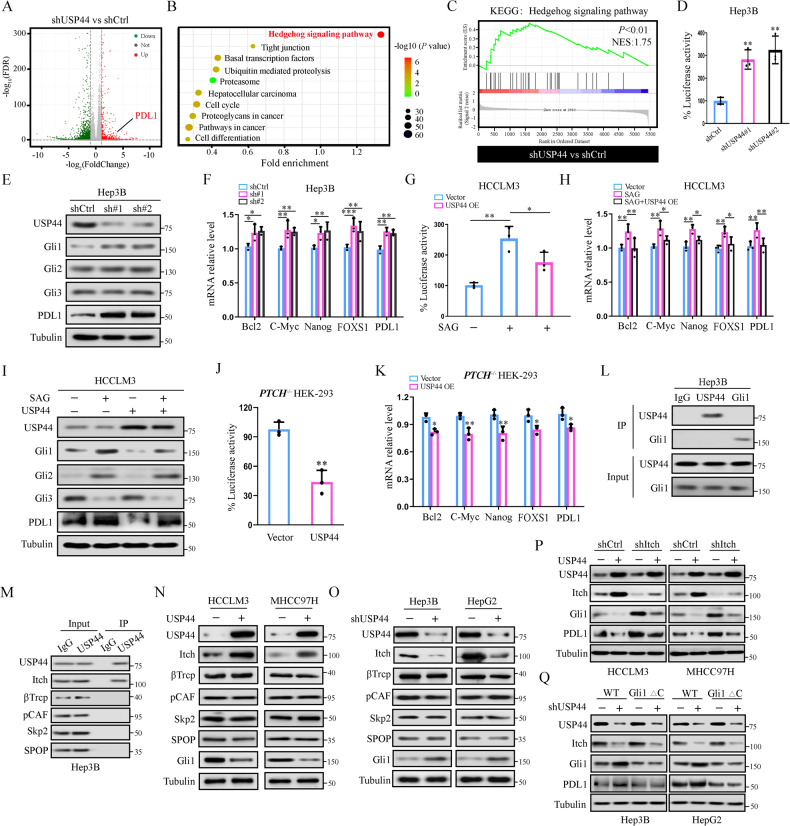
Hepatocellular carcinoma (HCC) is one of the deadliest malignancies in the world. Research into the key genes that maintain the malignant behavior of cancer cells is crucial for the treatment of HCC. Here, we identified ubiquitin-specific peptidase 44 (USP44), a member of the deubiquitinase family, as a novel regulator of HCC progression. The tumor suppressive function of USP44 was evaluated in a series of in vitro and in vivo experiments. Through quantitative proteomics examination, we demonstrated that USP44 inhibits HCC PDL1 expression by downregulating the Hedgehog (Hh) signaling pathway. Mechanistically, we found that USP44 directly interacts with Itch, an E3 ligase involved in Hh signaling, and promotes the deubiquitination and stabilization of Itch. These events result in the proteasomal degradation of Gli1 and subsequent inactivation of Hh signaling, which ultimately suppresses PDL1 expression and the progression of HCC. Furthermore, the HCC tissue microarray was analyzed by immunohistochemistry to evaluate the pathological relevance of the USP44/Itch/Gli1/PDL1 axis. Finally, the Gli1 inhibitor GANT61 was found to act in synergy with anti-PDL1 therapy. Overall, USP44 can act as a suppressive gene in HCC by modulating Hh signaling, and co-inhibition of Gli1 and PDL1 might be an effective novel combination strategy for treating HCC patients.


Conflict of interest statement

The authors declare no competing interests.

Figures

Fig. 1
Fig. 1. USP44 is downregulated and associated with a poor prognosis in HCC.
A Kaplan–Meier analysis of the overall survival and recurrence-free survival of HCC patients from the TCGA database. B Representative images of USP44 expression in paired tumors and peritumoral tissues from 14 patients with HCC using western blot. C mRNA expression of USP44 in 56-paired HCC tissues. D, E Low expression of USP44 was significantly related to poor overall survival and high recurrence rate in HCC patients. F Illustration of the mouse model of DEN/CCl4-induced hepatocarcinogenesis in USP44 flox/flox conditional knockout (KO) mice and their USP44 flox/flox littermates. G Gross appearance of representative livers with tumors were obtained from the indicated groups and PCNA staining of the tumor tissues. Scale bar, 1 cm (left panel) and 50 μm (right panel). H, I Tumor number and maximal tumor size were calculated in the spontaneous tumor models. J The relative number of PCNA+ cells were counted. *p < 0.05, **p < 0.01, ***p < 0.001 as indicated.
Fig. 2
Fig. 2. USP44 inhibits HCC metastasis and progression in vitro and in vivo.
A, B USP44 expression in different HCC cell lines and normal liver cell line L02 examined by western blot and qRT-PCR. CF The outcomes of USP44 knockdown and overexpression were examined by western blot and qRT-PCR in HCC cells. GN The Colony formation assay and the CCK-8 assay were used to detect the effect of USP44 on HCC proliferation. O, P The transwell assay was used to detect the effect of USP44 on the migration and invasion activities of HCC cells. Q, R Subcutaneous tumor model and orthotopic xenograft models were constructed using indicated cells, and tumor weights were measured at the endpoint (n = 6). S Representative H&E staining images of the lungs (left) and the number of metastases per lung (right) in each group. Data are obtained from three independent biological replicates and are presented as mean ± SD. Scale bar, 200 μm. *p < 0.05, **p < 0.01, ***p < 0.001 as indicated.
Fig. 3
Fig. 3. USP44 overexpression inhibits Hh signaling and PDL1 expression through Itch.
A Volcano plots showing DEPs in the shUSP44 group compared with the control group in Hep3B cells. |log2FC | >1, p-value < 0.05. B, C KEGG pathway enrichment and gene set enrichment analysis (GSEA) results indicated that hyperactivation of Hh signaling was observed in shUSP44 HCC cells compared with the control group. D Luciferase activity of the Hh pathway-reporter in shCtrl or shUSP44 transfected Hep3B cells. E Western blot for protein expression of Gli1, Gli2, Gli3, and PDL1 in shCtrl or shUSP44 transfected HCC cells. F mRNA level of Hh signaling target genes in shCtrl or shUSP44 transfected Hep3B cells. G Luciferase activity of the Hh pathway-reporter in control vector or exogenous USP44-overexpressing HCCLM3 cells treated with or without SAG (200 nM for 24 h). H mRNA level of Hh signaling target genes in control vector or exogenous USP44-overexpressing HCCLM3 cells treated with or without SAG (200 nM for 24 h). I Western blot for protein expression of Gli1, Gli2, Gli3, and PDL1 in control vector or exogenous USP44-overexpressing HCCLM3 cells treated with or without SAG (200 nM for 24 h). J Luciferase activity of the Hh signaling pathway-reporter in Ptch−/−HEK-293 cells expressing control vector or exogenous USP44. K mRNA level of Hh signaling target genes in Ptch−/−HEK-293 cells expressing control vector or exogenous USP44. L Interaction between USP44 and Gli1 was detected by immunoprecipitation followed by western blot analysis with the indicated antibodies. M Immunoprecipitation was employed to test the interaction between USP44 and the ubiquitin E3 ligases of Gli1. N, O Immunoblot for protein expression of the ubiquitin E3 ligases of Gli1 in HCC cells with different USP44 expression patterns. P Western blot for protein expression of Gli1 in HCC cells expressing control vector or exogenous USP44, with or without shItch transfection. Q Western blot for protein expression of Itch and Gli1 in shCtrl or shUSP44 expressing HCC cells transfected with wild-type Gli1 (Flag-Gli1 WT) or the mutant Gli1 (Flag-Gli1 ∆C). Data are obtained from three independent biological replicates and are presented as mean ± SD. *p < 0.05, **p < 0.01, ***p < 0.001 as indicated.
Fig. 4
Fig. 4. USP44 interacts with and deubiquitinates Itch.
A Western blot for protein expression of Itch in shCtrl or shUSP44 transfected HCC cells treated with or without MG132(15 μM, 6 h). B HCCLM3 cells were transfected with Flag-USP44 and Myc-Itch. Interaction between USP44 and Itch was detected by immunoprecipitation followed by western blot analysis with the indicated antibodies. C GST pull-down assays showed that GST-labeled Itch could directly interact with Flag-labeled USP44. D Co-localization of USP44 and Itch detected by immunofluorescence. Green and red, USP44 and Itch expressing cells, respectively. Nuclei were stained blue with DAPI. Scale bar indicates 10 μm. E shCtrl or shUSP44 transfected Hep3B cells were treated with CHX (20 μg/ml). The samples were collected at indicated time points and subjected to western blot analysis. The amounts of lysates from shCtrl and shUSP44 transfected Hep3B cells were adjusted to achieve similar levels of Itch at 0 h. F Control vector or exogenous USP44 transfected HCCLM3 cells were treated with CHX (20 μg/ml). The samples were collected at indicated time points and subjected to western blot analysis. The amounts of lysates from control vector and exogenous USP44 transfected HCCLM3 cells were adjusted to achieve similar levels of Itch at 0 h. G HA-Ub and wild-type or mutant USP44 were co-transfected into HCCLM3 cells, which were treated with 15 μM MG132 for 6 h before being harvested. The cells were immunoprecipitated with an anti-Itch antibody, followed by western blot analysis. H HA-Ub and shCtrl or shUSP44 were co-transfected into Hep3B cells, which were treated with 15 μM MG132 for 6 h before being harvested. The cells were immunoprecipitated with an anti-Itch antibody, followed by western blot analysis. I Deubiquitination of Itch in vitro by recombinant USP44. HEK-293T cells transfected with Myc-Itch and HA-Ub were treated with MG132 (15 μM) for 6 h before harvest. Cell lysates were incubated with FLAG Sepharose, then ubiquitinated Itch was incubated with or without purified recombinant USP44. The ubiquitin on Itch was analyzed by Western blot. Data are obtained from three independent biological replicates and are presented as mean ± SD. *p < 0.05, **p < 0.01, ***p < 0.001 as indicated.
Fig. 5
Fig. 5. USP44 impairs Hh signaling-dependent proliferation and migration of HCC cells in vitro.
A Itch and Gli1 protein expression in HCCLM3 cells expressing control vector or exogenous USP44, with or without Itch knockdown. B, C CCK-8 and EdU analyses for cell proliferation in HCCLM3 cells expressing control vector or exogenous USP44, with or without shItch transfection. Scale bar: 100 μm. D, E Transwell and wound-healing assays were used to test cellular invasion and migration in HCCLM3 cells expressing control vector or exogenous USP44, with or without shItch transfection. F Itch and Gli1 protein expression in control vector or exogenous USP44 expressing HCCLM3 cells transfected with wild-type Gli1 or the mutant Gli1. G, H CCK-8 and EdU analyses for cell proliferation in control vector or exogenous USP44 expressing HCCLM3 cells transfected with wild-type Gli1 or the mutant Gli1. Scale bar: 100 μm. I, J Transwell and wound-healing assays were used to test cellular invasion and migration in control vector or exogenous USP44 expressing HCCLM3 cells transfected with wild-type Gli1 or the mutant Gli1. Data are obtained from three independent biological replicates and are presented as mean ± SD. *p < 0.05, **p < 0.01, ***p < 0.001 as indicated.
Fig. 6
Fig. 6. USP44 inhibits Hh signaling-dependent growth and metastasis of HCC in vivo.
A Nude mice were subcutaneously injected with HCCLM3 cells stably expressing indicated plasmids. Intratumor protein levels of Itch and Gli1 in indicated groups were detected by western blot. B BALB/C nude mice subcutaneously injected with luciferase-expressing HCCLM3/Vector+shCtrl, HCCLM3/USP44+shCtrl, HCCLM3/Vector+shItch, or HCCLM3/USP44+shItch cells were assessed by IVIS imaging system. C, D Volume and weight of tumors in indicated groups (n = 5 per group). E IVIS imaging system was used to detect the effect of USP44 on metastasis of HCC in nude mice with orthotopic implanted xenografts. F Quantification of lung metastasis in the indicated groups of mice (n = 5 per group). G Nude mice were subcutaneously injected with HCCLM3 cells stably expressing indicated plasmids. Intratumor protein levels of Itch and Gli1 in indicated groups were detected by western blot. H Nude mice subcutaneously injected with luciferase-expressing HCCLM3/Vector, HCCLM3/USP44, HCCLM3/USP44+Gli1 WT, or HCCLM3/USP44+Gli1 MUT cells were assessed by IVIS imaging system. WT, wild-type; MUT, mutant. I, J Volume and weight of tumors in HCCLM3/Vector, HCCLM3/USP44, HCCLM3/USP44+Gli1 WT, or HCCLM3/USP44+Gli1 MUT groups. K IVIS imaging system was used to detect the effect of USP44 on metastasis of HCC in nude mice with orthotopic implanted xenografts. L Quantification of lung metastasis in the indicated groups of mice (n = 5 per group). Data are obtained from three independent biological replicates and are presented as mean ± SD. *p < 0.05, **p < 0.01, ***p < 0.001 as indicated.
Fig. 7
Fig. 7. GANT61 promotes the PDL1 therapy efficacy by targeting Gli1.
A, B Representative images and IHC score of IHC staining for indicated proteins in USP44-high and USP44-low HCC samples. Scale bar: 50 μm. C Correlations between USP44 and indicated protein levels detected from HCC tissues of 25 patients. D Schematic illustration of the therapy schedule for GANT61, anti-PDL1 mAb or combination therapy in orthotopic HCC mouse model using Hepa1-6 cell line. EG Representative images are presented at the endpoint and periodical detection of tumor development and progression using an ultrasound imaging platform (n = 6 per group). *p < 0.05, **p < 0.01, ***p < 0.001 as indicated.

References

    1. Sung H, Ferlay J, Siegel RL, Laversanne M, Soerjomataram I, Jemal A, et al. Global Cancer Statistics 2020: GLOBOCAN estimates of incidence and mortality worldwide for 36 cancers in 185 countries. CA Cancer J Clin. 2021;71:209–49. doi: 10.3322/caac.21660. - DOI - PubMed
    1. Finn RS, Ryoo BY, Merle P, Kudo M, Bouattour M, Lim HY, et al. Pembrolizumab as second-line therapy in patients with advanced hepatocellular carcinoma in KEYNOTE-240: a randomized, double-blind, phase III trial. J Clin Oncol. 2020;38:193–202. doi: 10.1200/JCO.19.01307. - DOI - PubMed
    1. Olatosi B, Vermund SH, Li X. Power of big data in ending HIV. AIDS. 2021;35:S1–S5. doi: 10.1097/QAD.0000000000002888. - DOI - PMC - PubMed
    1. Llovet JM, Ricci S, Mazzaferro V, Hilgard P, Gane E, Blanc JF, et al. Sorafenib in advanced hepatocellular carcinoma. N Engl J Med. 2008;359:378–90. doi: 10.1056/NEJMoa0708857. - DOI - PubMed
    1. Reyes-Turcu FE, Ventii KH, Wilkinson KD. Regulation and cellular roles of ubiquitin-specific deubiquitinating enzymes. Annu Rev Biochem. 2009;78:363–97. doi: 10.1146/annurev.biochem.78.082307.091526. - DOI - PMC - PubMed

Publication types

MeSH terms