. 2023 Dec 14;14(1):8191.
doi: 10.1038/s41467-023-43608-1.

Directing polymorph specific calcium carbonate formation with de novo protein templates

Affiliations

Directing polymorph specific calcium carbonate formation with de novo protein templates

Fatima A Davila-Hernandez et al. Nat Commun. .

Abstract

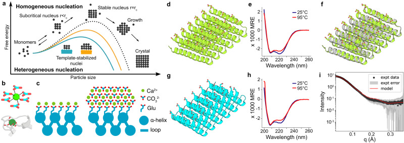
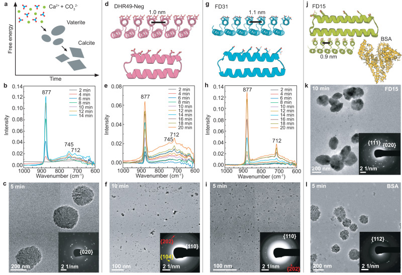
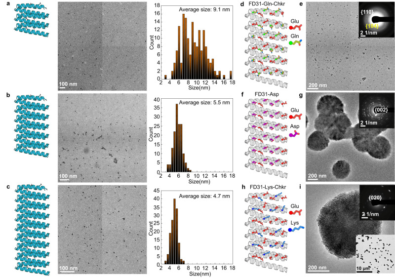
Biomolecules modulate inorganic crystallization to generate hierarchically structured biominerals, but the atomic structure of the organic-inorganic interfaces that regulate mineralization remain largely unknown. We hypothesized that heterogeneous nucleation of calcium carbonate could be achieved by a structured flat molecular template that pre-organizes calcium ions on its surface. To test this hypothesis, we design helical repeat proteins (DHRs) displaying regularly spaced carboxylate arrays on their surfaces and find that both protein monomers and protein-Ca2+ supramolecular assemblies directly nucleate nano-calcite with non-natural {110} or {202} faces while vaterite, which forms first in the absence of the proteins, is bypassed. These protein-stabilized nanocrystals then assemble by oriented attachment into calcite mesocrystals. We find further that nanocrystal size and polymorph can be tuned by varying the length and surface chemistry of the designed protein templates. Thus, bio-mineralization can be programmed using de novo protein design, providing a route to next-generation hybrid materials.


Conflict of interest statement

The authors declare no competing interests.

Figures

Fig. 1
Fig. 1. Design principles for the heterogeneous nucleation of CaCO3 guided by a protein template.
a The presence of a template reduces the free energy barrier of nucleation and the critical radius of the nuclei. b Coordination of calcium (top) by carbonate ions within a unit cell of calcite, and (bottom) by carboxylate containing glutamate and aspartate residues in the structure of calmodulin (PDB ID: 1A29, residues 18–33). c Tessellating binding moieties across repeated α-helices within a designed protein capable of pre-organizing bound calcium ions. di Biophysical characterization of the designed proteins. Design models for (d) FD15 and (g) FD31. e, h Their respective circular dichroism scans showing mean residue ellipticity (M.R.E.) from 200 to 260 nm at 25 °C and 95 °C. f The 4 Å resolution crystal structure of FD15 in gray (pdb id: 8UGC) superimposed on the design model in green (RMSD of 0.45 Å). i Experimental and model SAXS profiles for FD31, scattering vector (q, from 0 to 0.35 Å−1) vs. intensity.
Fig. 2
Fig. 2. Designed proteins modulate CaCO3 crystallization.
The CaCO3 crystallization process in supersaturated solutions containing 5 mM CaCl2 and 5 mM NaHCO3 was monitored in the absence and presence of different designed proteins. a Schematic showing the nucleation and transformation of CaCO3 in the absence of additives under the conditions used here. First, vaterite forms followed by transformation to micron-sized calcite. b In situ ATR-FTIR and c TEM of CaCO3 crystallization in the absence of protein showing the formation of vaterite. dl Impact of 1.08 μM designed proteins on mineralization. Protein design models (d, g) and corresponding in situ ATR-FTIR (e, h) show the formation of predominantly calcite in the presence of DHR49-Neg, and exclusively calcite in the presence of FD31. Representative TEM images confirm the formation of (c) vaterite in the absence of the proteins, f primarily calcite crystals with DHR49-Neg, and i calcite nanocrystals in the presence of FD31. In contrast, in the presence of (j) FD15 or a BSA control (j) only vaterite is formed (k, l). The TEM experiments in (c, f, i, k, l) were independently repeated three times with similar results.
Fig. 3
Fig. 3. FD31 mediated nucleation of calcite nanocrystals.
a Sequential LP-TEM images show the nucleation of eight CaCO3 nanoparticles (marked by yellow arrows) in a precursor solution containing FD31 protein. b Low-mag LP-TEM image shows more nucleated nanoparticles. c Sequential LP-TEM images show the initial nucleation and growth of two CaCO3 nanoparticles around the sheet-like templates after FD31 was preincubated with CaCl2 for 5 min before the addition of NaHCO3. Inset at t0 + 0 s displays multiple template formations at an early stage. A schematic (Inset at t0 + 13 s) shows the disconnected interface with a width of ≈ 1 nm between the as-formed particle and the substrate. At t0 + 129 s, the protein assembly dissolves, likely due to depletion of Ca, and the protein disperses into solution. Inset at t0 + 129 s shows an LP-HR-TEM image of a newly formed particle with the {110} lattice spacing of calcite. d In situ LP-HR-TEM image reveals the interface structure between calcite and the template. The yellow dotted lines highlight the interface. The FFT image in the inset shows the nucleated particle is calcite. As in Fig. 2g–i, the concentration of FD31 protein is 1.08 μM and CaCl2 and NaHCO3 is 5 mM. The TEM experiments were independently repeated three times with similar results.
Fig. 4
Fig. 4. Calcite nanocrystal growth and assembly in the presence of FD31.
a Sequential LP-TEM image sequence shows the attachment of multiple calcite nanocrystals into a larger one. b Ex situ TEM image showing calcite grown through particle attachment. The mean measured particle size of the 12 particles shown is 22.8 ± 3.9 nm. c HR-TEM image showing the aggregated crystal with {110} lattice orientation. White frames mark several individual calcite nanocrystals around the larger particle. d HR-TEM image shows additional calcite nanocrystals with a size of ≈ 20 nm and {110} side faces. The inset shows the calcite morphology and facet information. e TEM image shows ≈ 100 nm rhombohedral calcite. The inset FTT shows the crystal orientation and single crystallinity. Yellow and white arrows represent <104> and <110> growth orientations, respectively. f TEM image showing rhombohedral calcite with a size of ≈ 300 nm. The embedded SAED image demonstrates its single crystalline nature. Arrows mark several calcite nanocrystals on the surface. g HR-TEM and corresponding FFT images in the inset confirm the single crystalline nature and that the rough surface is composed of attached nanoparticles. Similar TEM results were collected in five independent experiments.
Fig. 5
Fig. 5. Tuning calcite nucleation by varying FD31 length and surface chemistry.
ac TEM and the size distribution of particles formed in the presence of a FD31-Rep3 with 3 repeats, b FD31 with 6 repeats, and c FD31-Rep9 with 9 repeats. The corresponding particle number (n) and mean measured particle size (S) are: a n = 172, S = 9.1 ± 2.9 nm, b n = 150, S = 5.5 ± 0.9 nm, c n = 147, S = 4.7 ± 0.8 nm, respectively. All the measurements are based on the representative TEM images from the same sample. di Representative TEM and SAED images show the formation of (e) calcite-dominant nanocrystals in the presence of FD31-Gln-Checker (d); the formation of (g) vaterite as well as calcite (Supplementary Fig. 12c, d) in the presence of (f) FD31-Asp; and the formation of a (i) vaterite dominant phase in the presence of (h) FD31-Lys-Checker. Three independent TEM experiments were performed with similar results.

References

    1. Gordon LM, Joester D. Nanoscale chemical tomography of buried organic–inorganic interfaces in the chiton tooth. Nature. 2011;469:194–197. doi: 10.1038/nature09686. - DOI - PubMed
    1. Jackson AP, Vincent JFV, Turner RM. Comparison of nacre with other ceramic composites. J. Mater. Sci. 1990;25:3173–3178. doi: 10.1007/BF00587670. - DOI
    1. Mann S. Molecular recognition in biomineralization. Nature. 1988;332:119–124. doi: 10.1038/332119a0. - DOI
    1. Addadi L, Weiner S. Interactions between acidic proteins and crystals: stereochemical requirements in biomineralization. Proc. Natl. Acad. Sci. USA. 1985;82:4110–4114. doi: 10.1073/pnas.82.12.4110. - DOI - PMC - PubMed
    1. De Yoreo JJ, et al. Crystallization by particle attachment in synthetic, biogenic, and geologic environments. Science. 2015;349:aaa6760. doi: 10.1126/science.aaa6760. - DOI - PubMed

Publication types