. 2023 Dec 14;14(1):8339.
doi: 10.1038/s41467-023-43823-w.

The bacterial replication origin BUS promotes nucleobase capture

Affiliations

The bacterial replication origin BUS promotes nucleobase capture

Simone Pelliciari et al. Nat Commun. .

Abstract

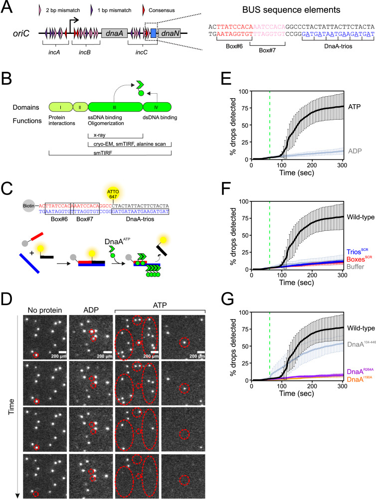
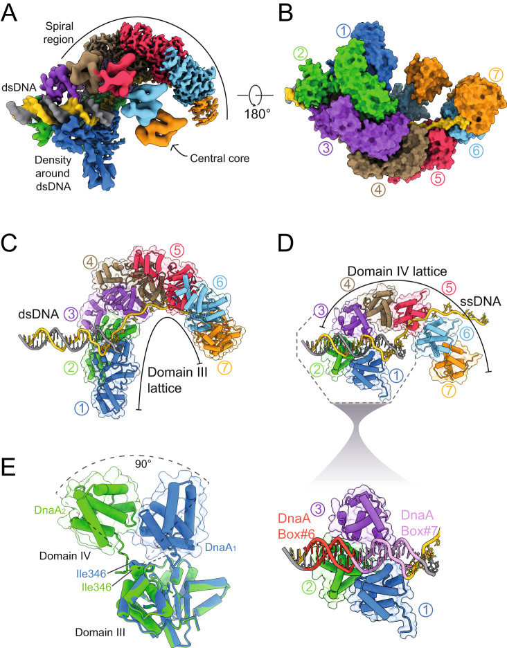
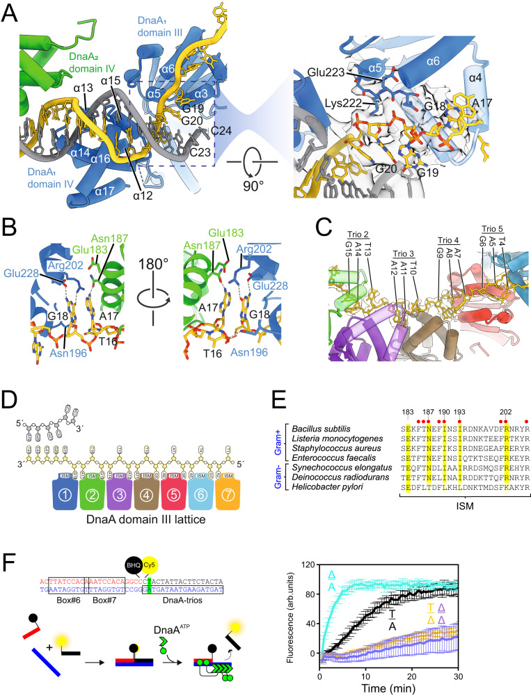
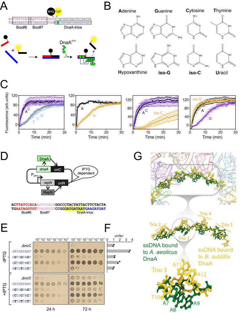
Genome duplication is essential for the proliferation of cellular life and this process is generally initiated by dedicated replication proteins at chromosome origins. In bacteria, DNA replication is initiated by the ubiquitous DnaA protein, which assembles into an oligomeric complex at the chromosome origin (oriC) that engages both double-stranded DNA (dsDNA) and single-stranded DNA (ssDNA) to promote DNA duplex opening. However, the mechanism of DnaA specifically opening a replication origin was unknown. Here we show that Bacillus subtilis DnaAATP assembles into a continuous oligomer at the site of DNA melting, extending from a dsDNA anchor to engage a single DNA strand. Within this complex, two nucleobases of each ssDNA binding motif (DnaA-trio) are captured within a dinucleotide binding pocket created by adjacent DnaA proteins. These results provide a molecular basis for DnaA specifically engaging the conserved sequence elements within the bacterial chromosome origin basal unwinding system (BUS).


Conflict of interest statement

The authors declare no competing interests.

Figures

Fig. 1
Fig. 1. DnaA strand separation events visualised by single molecule TIRF microscopy.
A Schematic representation of the B. subtilis replication origin and BUS sequence elements. DnaA-boxes are indicated with triangles. B Linear arrangement of DnaA domains I-IV. Locations of critical activities are shown. Endpoints of DnaA proteins used for various experiments are indicated. C Schematic describing single molecule TIRF microscopy experiments to investigate BUS activity. BUS sequence elements are indicated by colour. D Fields of view from single molecule TIRF experiments performed using different conditions (indicated on top) as a function of time. Red circles indicate spots that disappear over time. E Graph representing the percentage of fluorescent signals with decreased intensity over time in the presence of either ATP or ADP. F Graph reporting the percentage of fluorescent signals with decreased intensity over time using wild-type or mutant scaffolds. G Graph representing the percentage of fluorescent signals with decreased intensity over time using DnaA variants. DnaA104–446 lacks domains I-II. DnaAR264A is defective in oligomerisation. DnaAI190A is defective in ssDNA binding. For all graphs, data shows the average and percentage standard deviation from three independent experiments (source data are provided as a Source Data file).
Fig. 2
Fig. 2. Cryo-EM structure of the BUS complex and dsDNA engagement.
A Composite electron density map of the BUS, resulting from the assembly of maps corresponding to the spiral, central core, and the dsDNA regions contoured at 0.6σ, 0.21σ and 0.27σ respectively. The map is coloured based on seven DnaA protomers (blue, green, purple, brown, pink, cyan and orange respectively), the DnaA-trio containing DNA strand (yellow), and the complementary strand (grey). B Surface representation of the BUS complex coloured based on the respective DnaA protomers and DNA strands. C Model of the domain III lattice that makes up the spiral region of the map. Both the DNA scaffold and protein are shown in cylinder and stubs representation, with the protein also shown in a transparent surface representation. D Model of the domain IV lattice that makes up the central core of the map. Both the DNA scaffold and protein are shown in cylinder and stubs representation with the protein also shown in a transparent surface representation. The enlarged section shows domain IV of DnaA1 and DnaA2 engage with DnaA-box#7 and DnaA-box#6, respectively, while domain IV of DnaA3 engages the posterior minor groove. E Structural alignment of the DnaA1 and DnaA2 showing the domain IV of DnaA1 and DnaA2 are 90° apart relative to domain III.
Fig. 3
Fig. 3. Two bases of the DnaA-trio are flipped into the DnaA oligomer.
A Model showing engagement of α3 with the DNA scaffold and the loss of pairing between for G19:C24 and G20:C23. The focused image with a 90° rotated view shows residues Lys222 and Glu223 engaging with the phosphate groups of G20 and G19, respectively, together with the map density contoured at 0.6σ (spiral region map) and 0.34σ (dsDNA region map). B A focused view of the DnaA-trio#1 and DnaA-trio#3 interaction with DnaA domain III. Within the bipartite pocket G18 makes hydrogen bond interactions with Arg202 and Glu228 of DnaA1, while A17/A11 make hydrogen bond interactions with Asn187 and Glu183 of DnaA2 and DnaA4, respectively. Outside the bipartite binding pocket T16/T10 makes a hydrogen bond interaction with Asn196 of DnaA1 and DnaA3, respectively. C Interaction between the DnaA protomers and ssDNA, showing the two captured and one uncaptured base of DnaA-trio#2, #3, #4 and #5 with the extracted map around the bases contoured at 0.6σ. D Schematic showing the DnaA-trio strand engaging with DnaA protomers 1–7, where the first two bases of the DnaA-trio insert into the bipartite dinucleotide binding pocket while the third faces away. E Alignment of residues around the DnaA ISM involved in binding DnaA-trios (numbered for B. subtilis). Red dots indicate that alanine substitution in vivo is lethal. These DnaA homologs were previously shown to form a functional BUS with their cognate oriC sequences. F Schematic representation of the BHQ strand separation assay. BHQ-SSA performed using probes containing abasic sites (nucleotide substituted with THF) in the central position of the first DnaA-trio (base pair shown in green). Positions of bases on the top and bottom strand are indicated; Δ denotes an abasic site. Data shows the average and percentage standard deviation from three independent experiments (source data are provided as a Source Data file).
Fig. 4
Fig. 4. The amine group on adenine mediates specificity for DnaA-trio recognition by DnaA.
A Schematic representation of BHQ strand separation assay. The fluorescently labelled oligonucleotide contains an abasic site complementary to the central adenine (base shown in green). B Structures of nucleobases used to replace adenine. C Strand separation assay performed with nucleotide substitutions at the central adenine of the first DnaA-trio, in the context of an abasic site at the complementary position. Positions of mutations and structure of modified bases are indicated. Data shows the average and percentage standard deviation from three independent experiments (source data are provided as a Source Data file). D Schematic of the inducible repN/oriN system used to bypass mutations affecting oriC activity in B. subtilis. Replication via oriN is turned on and off in the presence and absence of IPTG, respectively. E Spot titre analysis with strains that have replaced the central adenine of DnaA-trio#1-3. The presence or absence of IPTG indicates the induction state of the repN/oriN system. Wild-type (FDS682), ΔincC (FDS404), A→G (FDS688), A→C (FDS686), A→T (FDS691). F Marker frequency analysis of strains shown in panel F. Data shows the average and individual data points from two independent experiments (source data are provided as a Source Data file). G Comparison of ssDNA substrates from DnaA nucleoprotein complexes. The synthetic polyA ssDNA from A. aeolicus (shown in green) (PDB 3R8F) was superimposed onto the BUS ssDNA (shown in yellow). Insets provide greater detail of the distinct conformations adopted by the two ssDNA substrates.

References

    1. O’Donnell, M., Langston, L. & Stillman, B. Principles and concepts of DNA replication in bacteria, archaea, and eukarya. Cold Spring Harb. Perspect. Biol.5 (2013). - PMC - PubMed
    1. Bleichert, F., Botchan, M. R. & Berger, J. M. Mechanisms for initiating cellular DNA replication. Science355, eaah6317 (2017). - PubMed
    1. Fernandez AJ, Berger JM. Mechanisms of hexameric helicases. Crit. Rev. Biochem. Mol. Biol. 2021;56:621–639. doi: 10.1080/10409238.2021.1954597. - DOI - PMC - PubMed
    1. Bell, S. P. & Kaguni, J. M. Helicase loading at chromosomal origins of replication. Cold Spring Harb. Perspect. Biol.5 (2013). - PMC - PubMed
    1. Messer W, et al. Functional domains of DnaA proteins. Biochimie. 1999;81:819–825. doi: 10.1016/S0300-9084(99)00215-1. - DOI - PubMed

Publication types