. 2024 Feb;31(2):150-158.
doi: 10.1038/s41418-023-01249-3. Epub 2023 Dec 14.

Genome-wide CRISPR screening identifies a role for ARRDC3 in TRP53-mediated responses

Affiliations

Genome-wide CRISPR screening identifies a role for ARRDC3 in TRP53-mediated responses

John E La Marca et al. Cell Death Differ. 2024 Feb.

Abstract

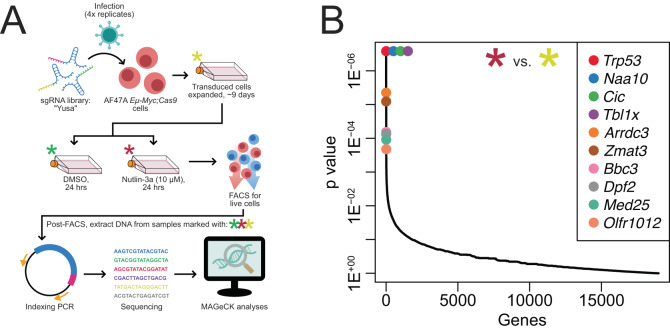
Whole-genome screens using CRISPR technologies are powerful tools to identify novel tumour suppressors as well as factors that impact responses of malignant cells to anti-cancer agents. Applying this methodology to lymphoma cells, we conducted a genome-wide screen to identify novel inhibitors of tumour expansion that are induced by the tumour suppressor TRP53. We discovered that the absence of Arrestin domain containing 3 (ARRDC3) increases the survival and long-term competitiveness of MYC-driven lymphoma cells when treated with anti-cancer agents that activate TRP53. Deleting Arrdc3 in mice caused perinatal lethality due to various developmental abnormalities, including cardiac defects. Notably, the absence of ARRDC3 markedly accelerated MYC-driven lymphoma development. Thus, ARRDC3 is a new mediator of TRP53-mediated suppression of tumour expansion, and this discovery may open new avenues to harness this process for cancer therapy.


Conflict of interest statement

The authors declare no competing interests.

Figures

Fig. 1
Fig. 1. Arrdc3 identified as a hit in a CRISPR/Cas9 screen for regulators of TRP53-mediated lymphoma growth suppression.
A Diagram of the CRISPR screen methodology using the MDM2 inhibitor nutlin-3a, which activates TP53/TRP53 in a non-genotoxic manner, as a selection pressure. In quadruplicate, AF47A Eμ-Myc lymphoma cells were transduced with the whole-genome “Yusa” sgRNA library, expanded, and either cultured without treatment or treated with either DMSO (vehicle control) or nutlin-3a (10 μM) for 24 h. Live cells were then sorted by FACS, DNA extracted from each sample (marked by asterisks), indexing PCR performed, and NGS combined with bioinformatic analyses used to identify sgRNAs that conferred a survival/competitive advantage after TRP53 activation. B Top hits from the CRISPR screen, comparing the untreated control cells (yellow asterisk) with the cells treated with nutlin-3a (red asterisk). Arrdc3 was identified as one of the top hits, with Arrdc3 targeting sgRNAs highly enriched in the surviving nutlin-3a-treated lymphoma cells.
Fig. 2
Fig. 2. Arrdc3 was validated as a TRP53 regulated gene and loss of Arrdc3 provided a competitive advantage to Eμ-Myc lymphoma cells after TRP53 activation.
A qRT-PCR analysis to assess the levels of Arrdc3 expression in AF47A Eμ-Myc lymphoma cells after treatment for 6 or 24 h with nutlin-3a (10 μM) or etoposide (40 ng/mL), relative to expression in DMSO-treated control cells. Additionally, expression of Pmaip1, Bbc3, and Cdkn1a were assessed as controls, and the expression of each gene was also examined in Trp53KO cells to assess the level of reliance of Arrdc3 expression on TRP53 activity. Gapdh was used as a housekeeping gene. Each treatment was performed three times, and the qRT-PCR was undertaken with 3 technical replicates. Error bars represent standard error of the mean. Statistical tests were one-way ANOVAs with Šídák’s multiple comparisons tests, performed to compare the indicated samples. Note that due to variation between biological replicates, some differences are not statistically significant despite obvious trends. B Cell cycle assay using the AF47A Eμ-Myc lymphoma cell line with CRISPR/Cas9-mediated knockout of Arrdc3 compared to the NTsgRNA transduced control lymphoma line, treated with 5 μM nutlin-3a for 6 h. Each treatment was performed 3 times. Data are presented as means +/− standard deviation. Statistical analyses are given in Table S1. C Cell death assays using the AF47A Eμ-Myc lymphoma cell line edited using CRISPR/Cas9 to ablate Arrdc3, treated for 24 h with one of three apoptosis-inducing drugs: nutlin-3a and etoposide that act via TP53/TRP53, or thapsigargin that functions in a TP53/TRP53-independent manner. Each cell line treatment was performed 3 times, with 2 technical replicates each time. Data are presented as means +/− standard deviation. D Cell competition assays using the 560 Eμ-Myc lymphoma cell line with CRISPR/Cas9-mediated knockout derivative lines of either Arrdc3 or Trp53, or a non-targeting sgRNA (NTsgRNA). Mixed cell populations (1:1) were treated with sub-optimal doses of either nutlin-3a (1.5 µM, ~IC20) or thapsigargin (1 nM, ~IC90) over a period of 14 days. Each competition assay was performed twice for each cell line, with 2 technical replicates each time, with a representative example being shown. Data are presented as means +/− standard deviation.
Fig. 3
Fig. 3. Arrdc3 knockout mice are not viable, with a range of abnormalities evident at embryonic day E19.5.
A The coding region of the Arrdc3 gene spans the region 81,031,503…81,044,161 on chromosome 13 in mice. Arrdc3 knockout mice were generated using CRISPR/Cas9 gene editing, with sgRNAs (red) targeting the 3’ UTR upstream of the gene and between exons (purple) 7 and 8, resulting in the deletion of the majority of the Arrdc3 gene locus. B The genotypes of offspring of inter-crosses between Arrdc3+/− mice do not obey Mendelian ratios post-birth (X2 = 82.87, df = 2, p < 0.05 (p < 1 × 10−15)), with only 8 Arrdc3−/− offspring being observed among 302 animals, none of which survived past weaning. By contrast, E14.5 foetuses generated from inter-crosses of Arrdc3+/− mice were seen to obey Mendelian ratios (X2 = 0.53, df = 2, p > 0.05 (p = 0.766)), with 31 of 137 foetuses genotyped as Arrdc3−/−. E18.5/19.5 pups also obeyed Mendelian ratios for each Arrdc3 genotype (X2 = 1.511, df = 2, p > 0.05 (p = 0.470)). Expected numbers are marked with a red E. C E19.5 Arrdc3−/− pups (n = 7 animals examined across 3 litters) displayed numerous gross morphological defects, though none were completely penetrant. D Underdeveloped eyes were observed in some Arrdc3−/− (and Arrdc3+/−) animals. In this stronger example, the right eye of mouse 470.3 was not externally visible. E An omphalocele observed in Arrdc3−/− mouse 468.3. F Haemorrhaging was observed on the exterior of some Arrdc3−/− animals and in some internal organs, including the surface of the thymus in Arrdc3−/− mouse 472.5. G Many Arrdc3−/− pups had breathing difficulties, including mouse 468.2, which also displayed additional morphological defects, including subcutaneous oedema which included serous fluid and blood.
Fig. 4
Fig. 4. Loss of Arrdc3 markedly accelerated MYC-driven lymphomagenesis in mice.
A Offspring of inter-crosses between Arrdc3+/− and Eμ-MycT/+;Arrdc3+/− mice do not obey Mendelian ratios post-birth (X2 = 122.11, df = 5, p < 0.05 (p < 1 × 10−15)). By contrast, E14.5 foetuses generated from such inter-crosses were seen to obey Mendelian ratios (X2 = 4.95, df = 5, p > 0.05 (p = 0.422)). Expected numbers are marked with a red E. B Kaplan–Meier survival curve of Eμ-MycT/+;Arrdc3+/+ mice (median survival = 91 days), Eμ-MycT/+;Arrdc3+/− mice (median survival = 77 days), and only one Eμ-MycT/+;Arrdc3−/− mouse that survived the developmental perinatal lethality. The absence of one allele of Arrdc3 slightly, but not significantly, accelerated lymphoma development in Eμ-Myc mice (Mantel-Cox test, df = 1, p > 0.05 (p = 0.0686)). C Kaplan–Meier lymphoma-free survival curve of lethally irradiated recipient mice that had been transplanted with Eμ-MycT/+;Arrdc3+/+ or Eμ-MycT/+;Arrdc3−/− foetal liver cells. We observed a statistically significant acceleration of lymphoma development in mice that had been transplanted with Eμ-MycT/+;Arrdc3−/− foetal liver cells (median survival = 67 days) compared to control mice that had been transplanted with Eμ-MycT/+;Arrdc3+/+ foetal liver cells (median survival = 210 days) (Mantel-Cox test, df = 1, X2 = 13.22, p < 0.001 (p = 0.000276)).

References

    1. Lossi L. The concept of intrinsic versus extrinsic apoptosis. Biochem J. 2022;479:357–84. - PubMed
    1. Nakano K, Vousden KH. PUMA, a novel proapoptotic gene, is induced by p53. Mol Cell. 2001;7:683–94. - PubMed
    1. Oda E, Ohki R, Murasawa H, Nemoto J, Shibue T, Yamashita T, et al. Noxa, a BH3-only member of the Bcl-2 family and candidate mediator of p53-induced apoptosis. Science. 2000;288:1053–8. - PubMed
    1. Tong T, Ji J, Jin S, Li X, Fan W, Song Y, et al. Gadd45a expression induces Bim dissociation from the cytoskeleton and translocation to mitochondria. Mol Cell Biol. 2005;25:4488–4500. - PMC - PubMed
    1. Yu J, Zhang L, Hwang PM, Kinzler KW, Vogelstein B. PUMA induces the rapid apoptosis of colorectal cancer cells. Mol Cell. 2001;7:673–82. - PubMed

Publication types