

CT texture features and lung shunt fraction measured using 99mTc-macroaggregated albumin SPECT/CT before trans-arterial radioembolization for hepatocellular carcinoma patients
- PMID: 38097801
- PMCID: PMC10721865
- DOI: 10.1038/s41598-023-49787-7
CT texture features and lung shunt fraction measured using 99mTc-macroaggregated albumin SPECT/CT before trans-arterial radioembolization for hepatocellular carcinoma patients
Abstract
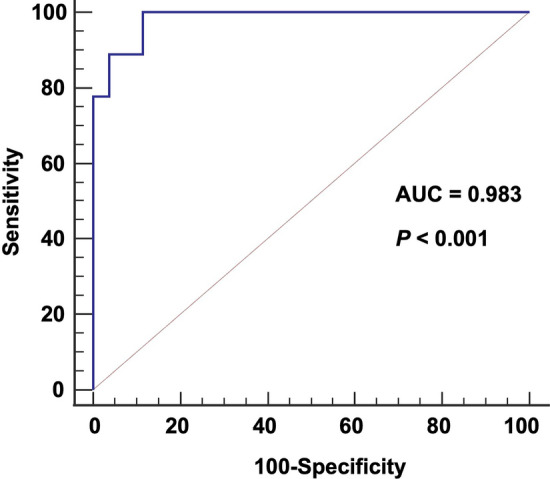
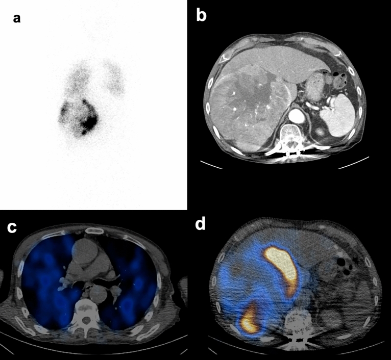
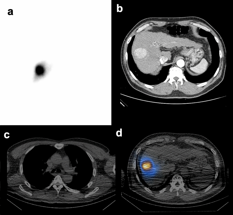
The aim of this study is to determine whether contrast-enhanced computed tomography (CECT)-based texture parameters can predict high (> 30 Gy) expected lung dose (ELD) calculated using 99mTc macroaggregated albumin single-photon emission computed tomography/computed tomography (SPECT/CT) for pre-trans-arterial radioembolization (TARE) dosimetry. 35 patients were analyzed, with a treatable planned dose of ≥ 200 Gy for unresectable hepatocellular carcinoma (HCC). Lung shunt fraction (LSF) was obtained from planar and SPECT/CT scans. Texture features of the tumor lesion on CECT before TARE were analyzed. Univariate and multivariate linear regression analyses were performed to determine potential ELD > 30 Gy predictors. Among the 35 patients, nine (25.7%) had ELD > 30 Gy, and had a higher LSF than the ELD ≤ 30 Gy group using the planar (20.7 ± 8.0% vs. 6.3 ± 3.3%; P < 0.001) and SPECT/CT (12.4 ± 5.1% vs. 3.5 ± 2.0%; P < 0.001) scans. The tumor integral total (HU × L) value was a predictor for high LSF using SPECT/CT, with an area under the curve, sensitivity, and specificity of 0.983 (95% confidence interval: 0.869-1.000, P < 0.001), 100%, and 88.5%, respectively. The tumor integral total value is an imaging marker for predicting ELD > 30 Gy. Applying CECT texture analysis may assist in reducing time and cost in patient selection and modifying TARE treatment plans.
© 2023. The Author(s).
Conflict of interest statement
The authors declare no competing interests.
Figures
Similar articles
-
Clinical and Dosimetric Implications of Calculating Lung Shunt Fraction for Hepatic 90Y Radioembolization Using SPECT/CT Versus Planar Scintigraphy.AJR Am J Roentgenol. 2022 Apr;218(4):728-737. doi: 10.2214/AJR.21.26663. Epub 2021 Oct 27. AJR Am J Roentgenol. 2022. PMID: 34704460
-
Radioembolization for Hepatocellular Carcinoma: a Comparison on Dual-phase Cone-beam CT, Contrast-enhanced CT (CECT) and 99mTc-macroaggregated albumin-SPECT/CT in predicting final distribution volumes and dosimetry of the post-embolization 90Y PET/CT.Radiol Med. 2025 Apr;130(4):474-485. doi: 10.1007/s11547-024-01946-0. Epub 2024 Dec 20. Radiol Med. 2025. PMID: 39707126 Free PMC article.
-
Optimizing dosimetric accuracy in radioembolization: comparative effectiveness of SPECT/CT, planar imaging, and PET/CT for lung shunt fraction assessment.Nucl Med Rev Cent East Eur. 2024;27(0). doi: 10.5603/nmr.102284. Nucl Med Rev Cent East Eur. 2024. PMID: 39726231
-
The role of SPECT/CT in radioembolization of liver tumours.Eur J Nucl Med Mol Imaging. 2014 May;41 Suppl 1:S115-24. doi: 10.1007/s00259-013-2675-5. Epub 2014 Jan 18. Eur J Nucl Med Mol Imaging. 2014. PMID: 24442600 Review.
-
Clinical impact of (99m)Tc-MAA SPECT/CT-based dosimetry in the radioembolization of liver malignancies with (90)Y-loaded microspheres.Eur J Nucl Med Mol Imaging. 2016 Mar;43(3):559-75. doi: 10.1007/s00259-015-3157-8. Epub 2015 Sep 4. Eur J Nucl Med Mol Imaging. 2016. PMID: 26338177 Free PMC article. Review.
References
-
- Garin E, et al. Personalised versus standard dosimetry approach of selective internal radiation therapy in patients with locally advanced hepatocellular carcinoma (DOSISPHERE-01): A randomised, multicentre, open-label phase 2 trial. Lancet Gastroenterol. Hepatol. 2021;6:17–29. doi: 10.1016/S2468-1253(20)30290-9. - DOI - PubMed
-
- Kennedy A, et al. Recommendations for radioembolization of hepatic malignancies using yttrium-90 microsphere brachytherapy: A consensus panel report from the radioembolization brachytherapy oncology consortium. Int. J. Radiat. Oncol. Biol. Phys. 2007;68:13–23. doi: 10.1016/j.ijrobp.2006.11.060. - DOI - PubMed
MeSH terms
Substances
LinkOut - more resources
Full Text Sources
Medical
