. 2023 Dec 14;15(1):114.
doi: 10.1186/s13073-023-01270-8.

Beyond the exome: utility of long-read whole genome sequencing in exome-negative autosomal recessive diseases

Affiliations

Beyond the exome: utility of long-read whole genome sequencing in exome-negative autosomal recessive diseases

Lama AlAbdi et al. Genome Med. .

Abstract

Background: Long-read whole genome sequencing (lrWGS) has the potential to address the technical limitations of exome sequencing in ways not possible by short-read WGS. However, its utility in autosomal recessive Mendelian diseases is largely unknown.

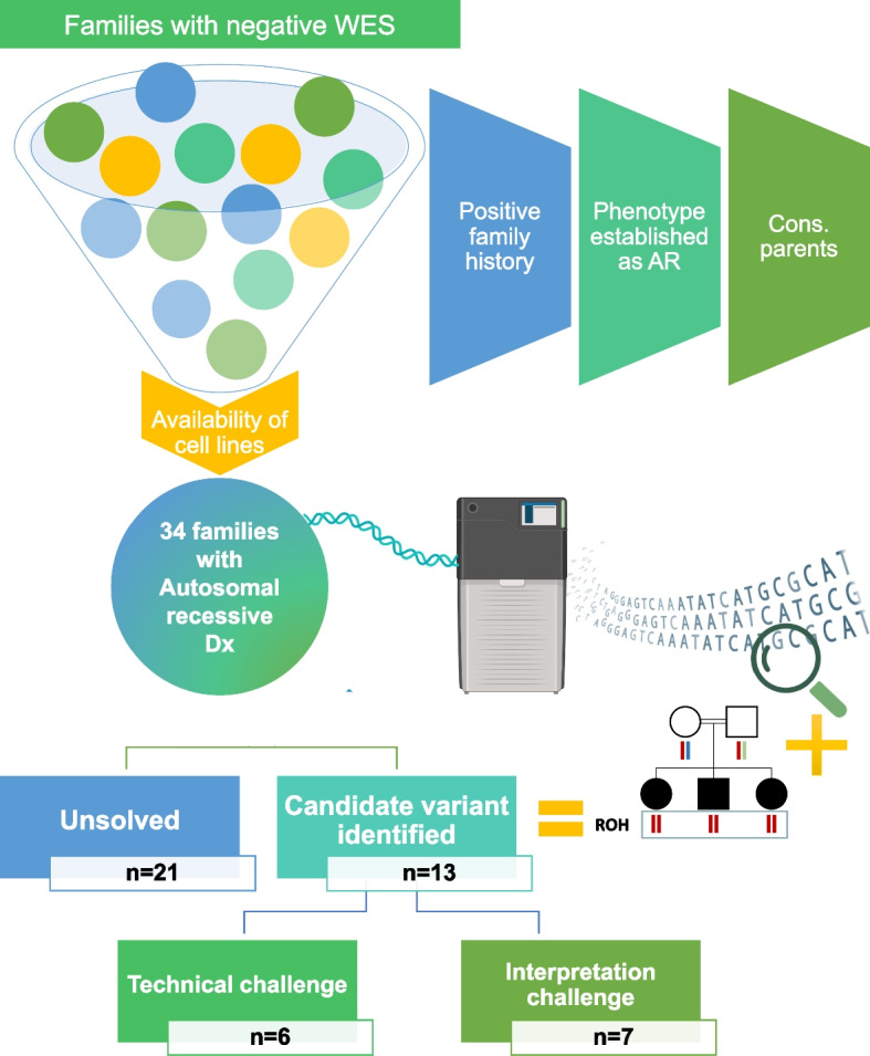
Methods: In a cohort of 34 families in which the suspected autosomal recessive diseases remained undiagnosed by exome sequencing, lrWGS was performed on the Pacific Bioscience Sequel IIe platform.

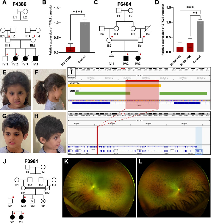
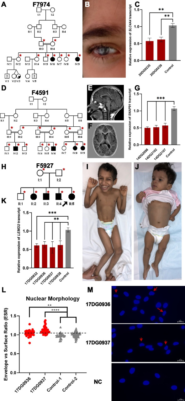
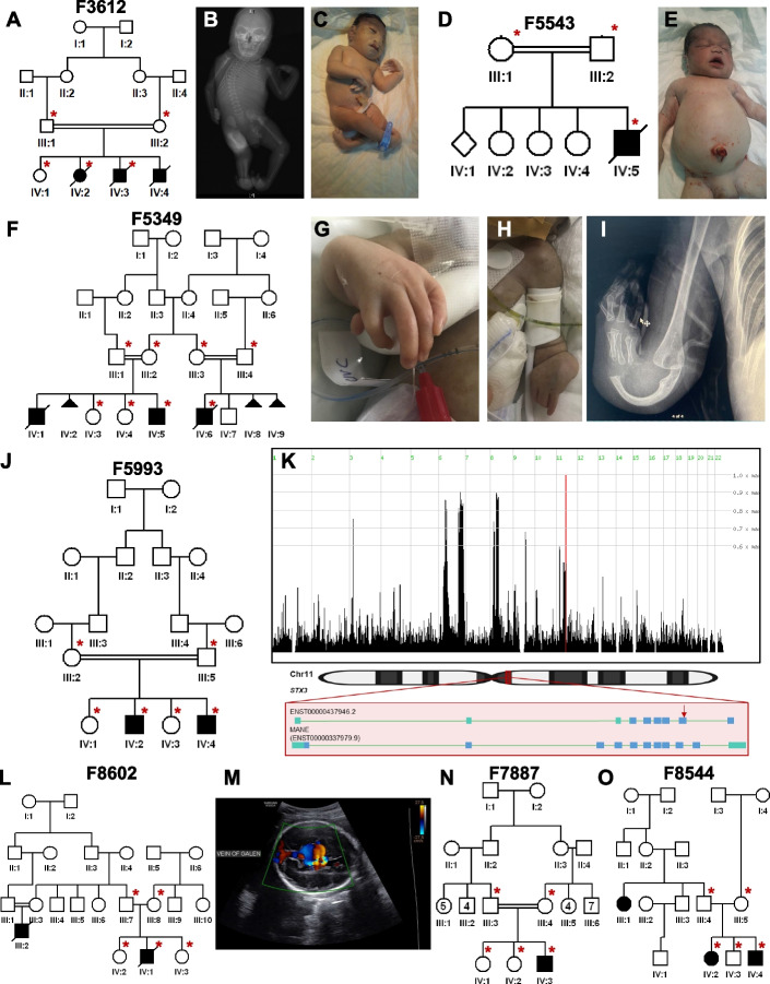
Results: Likely causal variants were identified in 13 (38%) of the cohort. These include (1) a homozygous splicing SV in TYMS as a novel candidate gene for lethal neonatal lactic acidosis, (2) a homozygous non-coding SV that we propose impacts STK25 expression and causes a novel neurodevelopmental disorder, (3) a compound heterozygous SV in RP1L1 with complex inheritance pattern in a family with inherited retinal disease, (4) homozygous deep intronic variants in LEMD2 and SNAP91 as novel candidate genes for neurodevelopmental disorders in two families, and (5) a promoter SNV in SLC4A4 causing non-syndromic band keratopathy. Surprisingly, we also encountered causal variants that could have been identified by short-read exome sequencing in 7 families. The latter highlight scenarios that are especially challenging at the interpretation level.

Conclusions: Our data highlight the continued need to address the interpretation challenges in parallel with efforts to improve the sequencing technology itself. We propose a path forward for the implementation of lrWGS sequencing in the setting of autosomal recessive diseases in a way that maximizes its utility.

Keywords: ABHD12; Autozygome; C1orf109; FLVCR1; Long-read sequencing; NID1; PKHD1; SHFM; STX3.


Conflict of interest statement

The authors declare that they have no competing interests.

Figures

Fig. 1
Fig. 1
Schematic presentation of the study design. All cases with negative exome reanalysis with available cell lines were considered if they met at least two of the following criteria: the case has a positive family history for the same disease, the phenotype is established as autosomal recessive, and the parents are consanguineous. Ultramolecular weight DNA was extracted from established cell lines for each case and underwent lrWGS using PacBio technology. Autozygome-guided analysis was performed, and candidate variants were identified in 13 families. Six variants were not identified by exome or its reanalysis and presented technical challenges while the remaining seven variants presented interpretation challenges. Cons: consanguineous. Some illustrations were created using BioRender.com
Fig. 2
Fig. 2
Structural variants that are not detected by exome. A Pedigree of family F4386 with three children who died as neonates with lactic acidosis. B RT-qPCR results showing reduced transcript levels of TYMS in one of the affected individuals compared to two independent controls. C Pedigree of family F6404 with two siblings affected with microcephaly, and developmental delay. D RT-qPCR data showing reduced expression of STK25 in the two affected individuals compared to two independent control samples. EH clinical images of the two siblings highlighting the microcephaly and lack of gross facial dysmorphism. E and F Clinical images of 20DG0785. G and H Clinical images of 20DG0786. I Genomic representation of the deletion with H3K27Ac and DNase hypersensitivity signals indicated. J Pedigree of family F3981 with two affected sisters with Leber congenital amaurosis and their affected mother with retinitis pigmentosa. K and L Widefield retinal imaging of the left and right retina of the mother (IV:2) showing rod cone dystrophy. Error bars denote standard deviation of at least 3 experiments. ****, ***, ** denote p-values < 0.0001, < 0.001, and < 0.01, respectively, using unpaired Student’s t-test
Fig. 3
Fig. 3
Challenging variants that are not detected by exome. A Pedigree of family F7974 with two sisters affected with band keratopathy. B Clinical images of individual (IV:6) highlighting keratopathy phenotype. C RT-qPCR experiment showing reduced SLC4A4 transcript levels compared to two independent controls. D Pedigree of family F4591 with three cousins affected with microcephalic NDD. E and F MRI imaging of individual (IV:8) showing mild brain atrophy and thin corpus callosum. G RT-qPCR results demonstrating reduced expression of SNAP91 in three patients compared to two independent controls. H Pedigree of family F5927 with four children affected with NDD. I and J Clinical images of individuals (II:3 and II:4) highlighting microcephaly and progressive spasticity. K RT-qPCR experiment showing consistently reduced LEMD2 expression levels in samples from four affected siblings compared to two independent controls. L and M Nuclear morphology of patient cells compared to two independent controls and representative images highlighting abnormal nuclear morphology using red arrows. Error bars denote standard deviation of at least 3 experiments. ****, ***, ** denote p-values < 0.0001, < 0.001, and < 0.01, respectively, using unpaired Student’s t-test
Fig. 4
Fig. 4
Families solved with variants that represented interpretation challenges. A Pedigree of family F3612 with three stillbirths all presenting with microcephaly and skeletal dysplasia phenotypes. B and C X-ray and clinical image of an affected individual (IV:4) highlighting radial ray deficiency. D Pedigree of family F5543 with an affected baby with polycystic kidney disease. E Clinical image of individual (IV:5) with Potter facies and swollen abdomen. F) Pedigree of family F5349 referred to us with two affected cousins both presenting with severe limb malformations. G and H Clinical images of affected individual (IV:6) with absent radius and hypoplasia of the ulna. I X-ray images of the right hand of affected individual (IV:6) highlighting absent radial ray and bowed ulna. J Pedigree of family F5993 with two brothers affected with retinal dystrophy. K (top) Genomic representation of identical haplotypes between two families homozygous for the same STX3 variant. HomozygosityMapper shows that they map to a single locus on Chr.13; (bottom) schematic representation of two STX3 transcripts, one where the variant is a frameshift insertion and the other (MANE select) as deep intronic. L Pedigree of family F8602 with two cousins affected with vein of Galen malformation. M Brain ultrasound imaging of individual (IV:2) showing dilatation of the veins of Galen. N Pedigree of family F7887 with NDD. O Pedigree of family F8544 with two siblings and a cousin affected with NDD. Illustrations at the bottom of panel (K) were created using BioRender.com
Fig. 5
Fig. 5
Variants that were not detected by lrWGS. A Pedigree of family F6440 with three stillbirths all presenting with profound global developmental delay, microcephaly, epilepsy, and scoliosis. B Clinical image of an affected individual (III:10) highlighting microcephaly. C EasyLinkage analysis showing a single locus at chr9:31537680–79306780 with LOD of 4. D Screenshot of Bionano analysis output identifying an insertion (chr9:33,266,774–33,271,717) disrupting CHMP5. E RT-PCR experiment showing 91 bp insertion in intron 2 (I2). F Western blot experiment showing reduction of CHMP5 protein levels in two affected individuals compared to two independent controls. Alpha-Tubulin was used as loading control. G Relative quantification of CHMP5 protein levels showing ~ 80% reduction compared to controls. H Pedigree of family F8280 with two siblings affected with SHFM. I Clinical image of limb malformation of affected individual (IV:1). J Chromosomal microarray output from family F8280 showing heterozygous duplication in the known SHFM locus that was absent in the parents

References

    1. Alkuraya FS. How the human genome transformed study of rare diseases. Nature. 2021;590:218–9. - PubMed
    1. Shickh S, Mighton C, Uleryk E, Pechlivanoglou P, Bombard Y. The clinical utility of exome and genome sequencing across clinical indications: a systematic review. Hum Genet. 2021;140(10):1403–1416. doi: 10.1007/s00439-021-02331-x. - DOI - PubMed
    1. Marwaha S, Knowles JW, Ashley EA. A guide for the diagnosis of rare and undiagnosed disease: beyond the exome. Genome medicine. 2022;14(1):1–22. doi: 10.1186/s13073-022-01026-w. - DOI - PMC - PubMed
    1. Lowther C, Valkanas E, Giordano JL, Wang HZ, Currall BB, O'Keefe K, et al. Systematic evaluation of genome sequencing for the diagnostic assessment of autism spectrum disorder and fetal structural anomalies. Am J Hum Genet. 2023;110(9):1454–1469. doi: 10.1016/j.ajhg.2023.07.010. - DOI - PMC - PubMed
    1. Investigators GPP, Smedley D, Smith KR, Martin A, Thomas EA, McDonagh EM, et al. 100,000 Genomes Pilot on Rare-Disease Diagnosis in Health Care - Preliminary Report. N Engl J Med. 2021;385(20):1868–1880. doi: 10.1056/NEJMoa2035790. - DOI - PMC - PubMed

Publication types

LinkOut - more resources