. 2023 Dec 14;20(1):93.
doi: 10.1186/s12987-023-00500-w.

Structural characterization of SLYM-a 4th meningeal membrane

Affiliations

Structural characterization of SLYM-a 4th meningeal membrane

Virginia Plá et al. Fluids Barriers CNS. .

Abstract

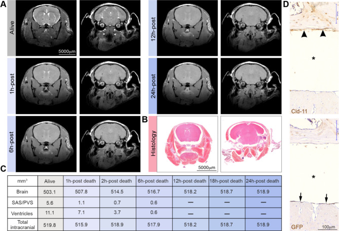
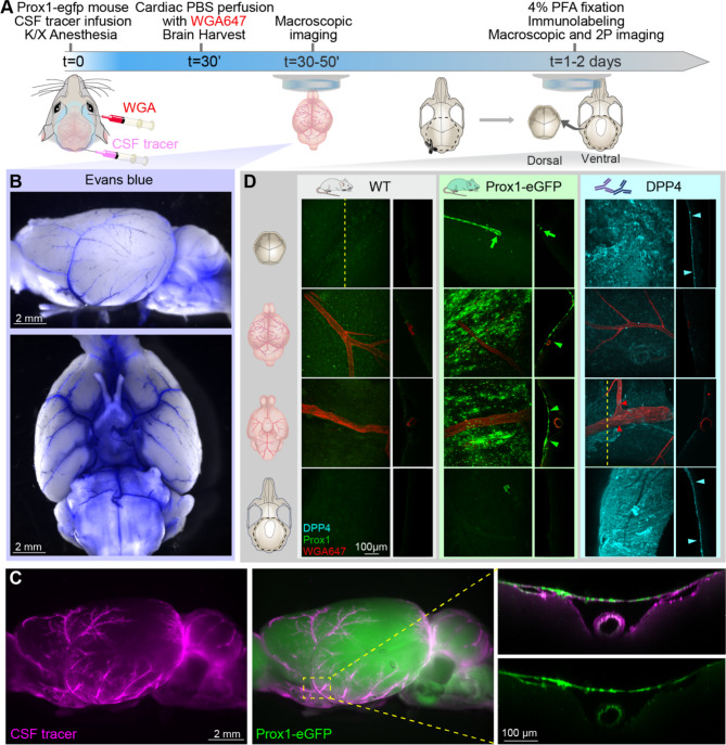
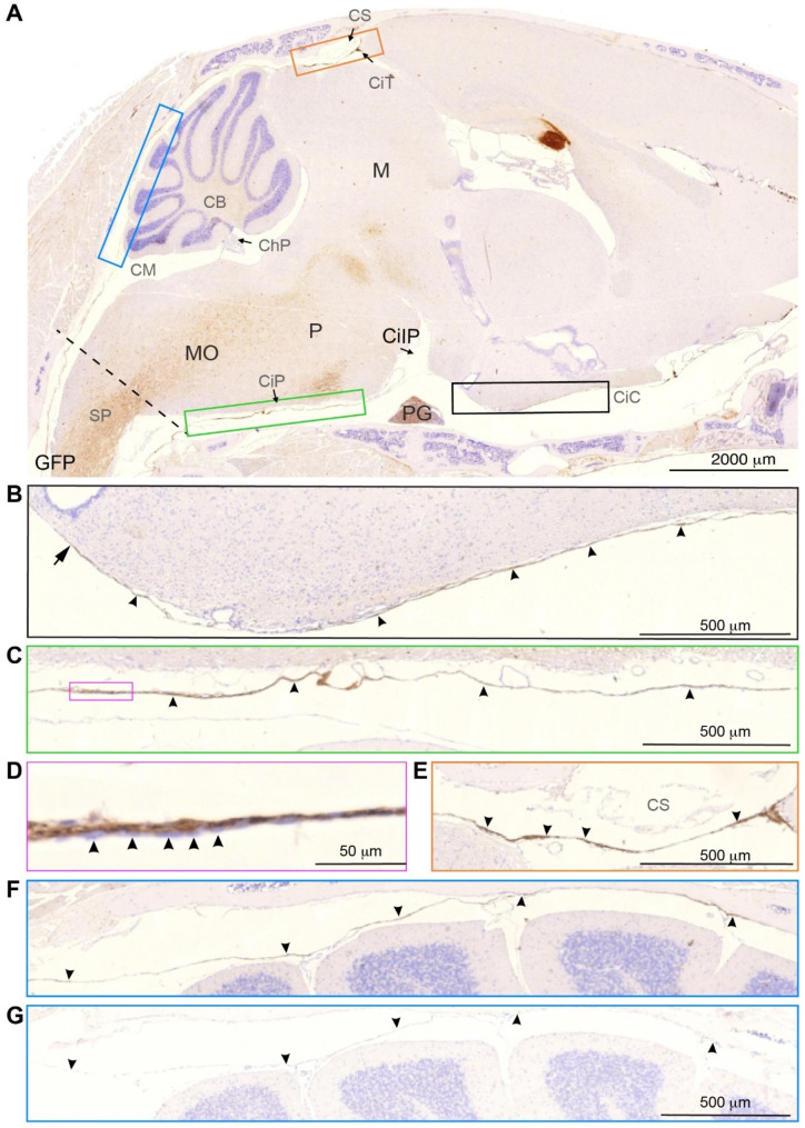
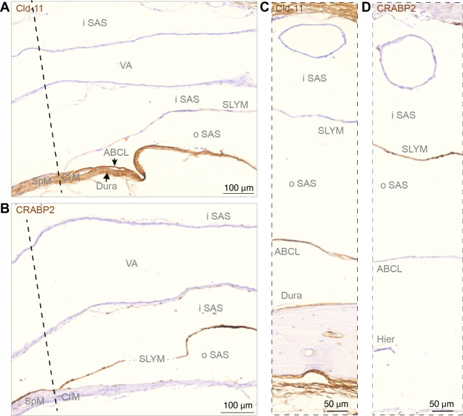
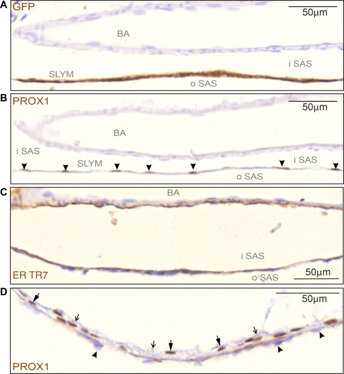
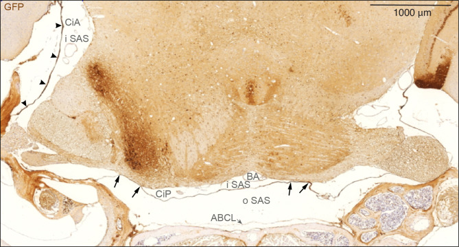
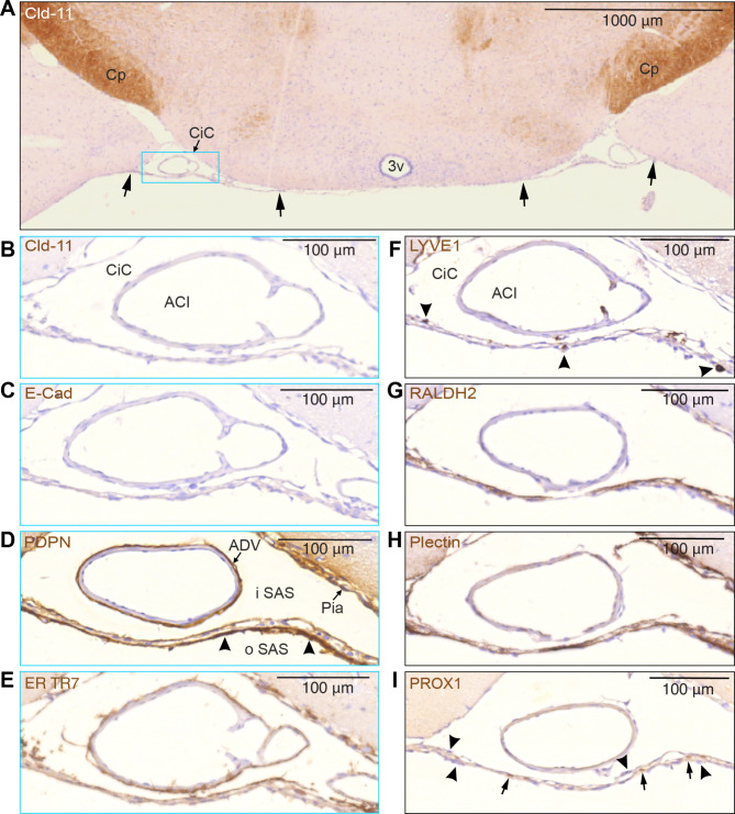
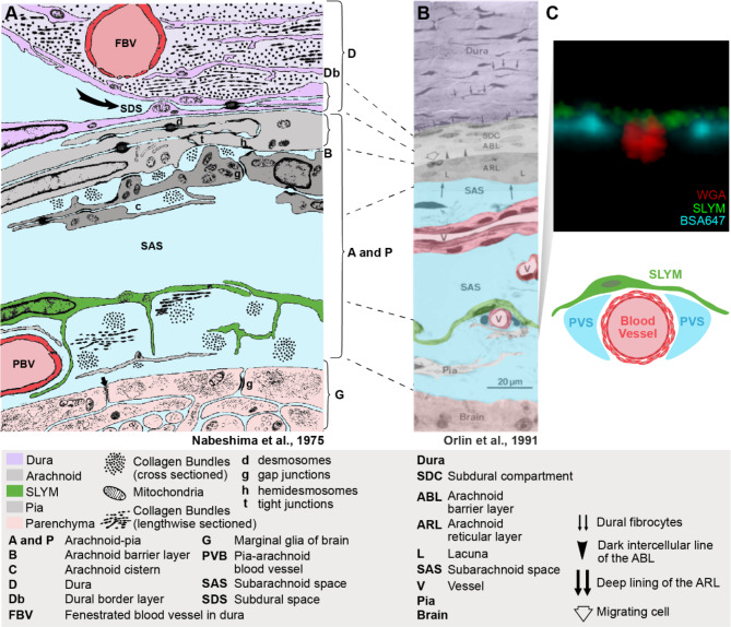
Traditionally, the meninges are described as 3 distinct layers, dura, arachnoid and pia. Yet, the classification of the connective meningeal membranes surrounding the brain is based on postmortem macroscopic examination. Ultrastructural and single cell transcriptome analyses have documented that the 3 meningeal layers can be subdivided into several distinct layers based on cellular characteristics. We here re-examined the existence of a 4th meningeal membrane, Subarachnoid Lymphatic-like Membrane or SLYM in Prox1-eGFP reporter mice. Imaging of freshly resected whole brains showed that SLYM covers the entire brain and brain stem and forms a roof shielding the subarachnoid cerebrospinal fluid (CSF)-filled cisterns and the pia-adjacent vasculature. Thus, SLYM is strategically positioned to facilitate periarterial influx of freshly produced CSF and thereby support unidirectional glymphatic CSF transport. Histological analysis showed that, in spinal cord and parts of dorsal cortex, SLYM fused with the arachnoid barrier layer, while in the basal brain stem typically formed a 1-3 cell layered membrane subdividing the subarachnoid space into two compartments. However, great care should be taken when interpreting the organization of the delicate leptomeningeal membranes in tissue sections. We show that hyperosmotic fixatives dehydrate the tissue with the risk of shrinkage and dislocation of these fragile membranes in postmortem preparations.

Keywords: Cerebrospinal fluid; Glymphatic system; Perivascular space; Prospero Homeobox 1; Subarachnoid space.


Conflict of interest statement

The authors declare no competing interests.

Figures

Fig. 1
Fig. 1
Preparation of histological sections is linked to a marked shrinkage of the brain’s fluid-filled spaces. (A) Magnetic resonance imaging (MRI) of a young isoflurane anesthetized C57BL/6JRj mouse. Bright white signal represents CSF in the subarachnoid space (SAS), perivascular spaces (PVS) and the ventricles which gradually disappears after death, indicative of brain swelling. After the live imaging session, the anesthetized mouse was killed by 100% nitrogen inhalation via a nose cone on an MR-compatible stereotactic holder, and scanning was repeated at 1, 6, 12, 18, and 24 h after cardiorespiratory arrest. (B) Histological section of the head of a young C57BL/6JRj mouse after decalcification and preparation of parafin. (C) Quantification of the volume of the brain, SAS/PVS, ventricles and total intracranial volume based on the images displayed in panel A. (D) Immunohistochemical analysis of arachnoid barrier cell layer (top, arrowheads) stained for Cld-11 and SLYM stained for GFP (lower, arrows) in two consecutive sections prepared from a Prox1-eGFP mouse. Note the considerable shrinkage of the brain (asterisk) in the sections
Fig. 2
Fig. 2
SLYM defines the subarachnoid cisternal system and the pial perivascular spaces. (A) Schematic of the experimental approach. CSF tracers, either Evans Blue (0.9 kDa, 0.5% in aCSF w/v) or BSA-647 (67 kDa, 0.5% in aCSF w/v) were delivered (10 µL, 2 µL/min) through a cisterna magna cannula in K/X-anesthetized Prox1-eGFP mice. After 30 min of tracer circulation, the Prox1-eGFP mice were perfused with phosphate buffered saline (PBS) to remove blood signal interference. In a subset of experiments, wheat germ agglutinin (WGA) conjugated to Alexa Fluor 647 was injected retro-orbitally to outline the vasculature. (B) Macroscopic fluorescent images of the whole brain showing the cisterns and pial perivascular spaces filled with a small size tracer, Evans blue (blue). (C) Representative images of a brain from a Prox1-eGFP (green, middle) mouse injected with a larger CSF tracer, BSA-647 (magenta, left). 2-photon images of middle cerebral artery (right) illustrate the accumulation of the fluorescent tracer below the Prox1-eGFP+ cells. The CSF tracer is taken up by cells bordering the perivascular spaces after death, including the arterial smooth muscle cells and pia [11]. (D) Confocal images of skull and brain from Prox1-eGFP and wildtype mice taken before and after DPP4 staining. Confocal and two-photon XY projections from whole-mount dorsal skull (first row), dorsal brain (second row), ventral brain (third row), and ventral skull (bottom row). Orthogonal projections were obtained as indicated by the dashed line in the first panel, except for the ventral brain after DPP4 staining where the projection was obtained from an area including a DPP4 positive section close to a vessel outline by WGA. A meningeal lymphatic vessel positive for Prox1-eGFP is observed in the dorsal skull of the Prox1-eGFP mouse (green arrow). The analysis showed that SLYM in the Prox1-eGFP reporter mice (green arrowheads) adheres to the brain surface, whereas the ABC layer, immunolabeled for DPP4 (cyan arrowheads), adheres to dura on the skull
Fig. 3
Fig. 3
SLYM encases the brain parenchyma from the upper spinal cord to the olfactory bulb region. (A) Sagittal section from a Prox1-eGFP mouse stained for GFP. GFP immunosignal forms a lamina covering the entire brain, delimiting the cisternal spaces. Colored insets are used to show high magnification images of different anatomical regions. The dashed line indicates the plane through foramen magnum separating the spinal cord (SP) from medulla oblongata (MO). (B) Black insert shows the rostral extension of the basal cistern (arrowheads). Note that the histochemical reactivity is attenuated corresponding to the median eminence (arrow). Cisterna interpeduncularis (CiIP) has been already analyzed in a previous publication (Møllgård, et al., 2023 [12], suppl. Fig. 3). (C) Green insert depicts the cisterna pontis (CiP), lining the basal CSF space. (D) Magenta insert shows a higher magnification view of SLYM, where individual GFP positive cells (arrowheads) seem to be partly covered by an inner cell layer to the left. (E) Orange insert shows the region of confluence of sinuses (CS) where the GFP signal is clearly visible (arrowheads), delimiting the cisterna tegmentalis (CiT). (F) Blue insert shows the superior cerebellar cistern (cisterna vermis), delimiting two CSF spaces adjacent to the cerebellum. The one-layered delicate SLYM membrane deviates between attachment to the skull and the cerebellar pial surface. (G) Blue insert depicts an adjacent section to panel F stained following omission of the primary (GFP antibody. The delicate unstained SLYM membrane is present but can barely be identified. Arrowheads are placed in the same positions in F and G. CB: cerebellum, ChP: choroid plexus, CiC: carotidand chiasmatic cistern, CiIP: cisterna interpeduncularis. CiP: cisterna pontis, CiI: cisterna tegmentalis, CM: cisterna magna, CS: confluence sinuum, M: mesencephalon, MO: medulla oblongata, P: pons, PG: pituitary gland, SP: spinal cord. Magnification is indicated by labelled bars on the individual figures
Fig. 4
Fig. 4
Sagittal sections show SLYM is not fused with the arachnoid barrier layer along basal cisterns. (A, B) Immunohistochemical analysis of arachnoid barrier cell layer (ABCL) stained for Cld-11 and SLYM stained for CRABP2 at the transition from spinal meninges (SpM) to cranial meninges (CeM) indicated by dashed line, corresponding to the plane of foramen magnum. SLYM and ABCL are fused corresponding to the rostral-most spinal meninges, but the deviation of the two layers is characteristic of the beginning of cranial meninges creating an inner subarachnoid space (iSAS) containing the vertebral artery (VA) and an outer subarachnoid space, between SLYM and the arachnoid barrier layer. In (C, D) corresponding to cisterna pontis the arachnoid barrier cell layer (ABCL), positively reacting for Cld-11, covers dura but renders SLYM negative. SLYM shows positive immunoreactivity for CRABP2 whereas the ABCL is not stained. Due to the heat-induced epitope retrieval (Hier) a fraction of bone with dura disappeared from this section. Dashed line indicates a nearly frontal plane through foramen magnum separating the spinal cord from medulla oblongata and thus the cranial meninges (CrM) from spinal meninges (SpM). ABCL: arachnoid barrier cell layer, CrM: cranial meninges, Hier: heat-induced epitope retrieval, iSAS: inner subarachnoid space, oSAS: outer subarachnoid space, SpM: spinal meninges, VA: vertebral artery. Magnification is indicated by labelled bars on the individual figures
Fig. 5
Fig. 5
SLYM is characterized by a specific subset of immunological markers. (AC) High magnification images of the basilar artery on the ventral part of the brain show the division of the subarachnoid space (SAS) into an outer and inner compartment by the SLYM meningeal layer (GFP + labelled). (B) Prox1 immunolabeling of serial sections of the same tissue allowed individual identification of SLYM cells, due to their immunoreactive nuclei (arrowheads). (C) ER-TR7 immunoreactivity is found on the inner surface of SLYM, showing the presence of Collagen type VI on this meningeal layer. Also note the staining of adventitia of the basilar artery (BA). (D) The Prox1-stained nuclei (thick arrows) in the wall of cisterna ambiens are in direct contact with arachnoid barrier cells towards the exterior (arrowheads) and with an unstained layer of arachnoid reticular cells facing the inner subarachnoid space (thin, open arrows). BA: basilar artery, iSAS: inner subarachnoid space, oSAS: outer subarachnoid space. Magnification is indicated by labelled bars on the individual figures
Fig. 6
Fig. 6
SLYM encases brain parenchyma evident in a coronal section through mid-pontine region. Immunohistochemical analysis of GFP signal of a coronal section of Prox1-eGFP tissue allowed additional characterization of the meningeal layers covering the parenchyma. SLYM (GFP+) layer divides the subarachnoid space into two well-defined compartments, an outer subarachnoid space (oSAS) facing the arachnoid barrier cell layer (ABCL) and an inner subarachnoid space (iSAS), enclosing blood vessels, here the basilar artery (BA). SLYM seems to fuse with pia covering a fraction of the basolateral part of pons on both sides (arrows) thus creating a sub-compartmentalization of the inner SAS. The strongly stained cisterna ambiens (CiA) is indicated by arrowheads. ABCL: arachnoid barrier cell layer, BA: basilar artery, CiA: cisterna ambiens, CiP: cisterna pontis, iSAS: inner subarachnoid space, oSAS: outer subarachnoid space. Magnification is indicated by labelled bar
Fig. 7
Fig. 7
SLYM and the ABC layer can be differentiated by their distinctive immunomarker expression. (A) The unstained SLYM in this claudin-11 stained section located at a middle coronal position of the circle of Willis, at the suprachiasmatic level, indicated by arrows, seems to fuse with the pia along both medial and lateral borders of the cisterna caroticus (CiC) -blue rectangle- which is shown in higher magnification in (BI). Note the positive Cld-11 reactivity in the oligodendrocytes of the cerebral peduncle (Cp) in contrast to the negative appearance of SLYM in (B). (C) E-cadherin staining also provided a negative response in SLYM, pia and adventitia of the internal carotid artery (ACI). (DI) show SLYM covered by an inner and outer layer of different types of arachnoid leptomeningeal fibroblasts. The PDPN-stained section (D) demonstrates a strongly reactive layer (arrowheads) facing the outer SAS (oSAS), a weaker stained SLYM in the middle and yet another thin leptomeningeal layer facing the inner SAS (iSAS) with a positively reacting pia. The adventitia (ADV) surrounding the internal carotid artery (ACI in B) is also positively stained. (E) ER-TR7 immunoreactivity demonstrates collagen type VI in particular on the inside of SLYM but is also associated with adventitia of ACI and pia. (F) As expected by its “lymphatic-like” nature, SLYM acts as an immunological niche. LYVE1 antibody highlights perivascular macrophages (arrowheads) located either on the inner, middle or outer part of SLYM. (G) Retinaldehyde dehydrogenase (RALDH2) is particularly strongly stained along the inner SLYM layer. (H) Plectin, a possible key cytoskeleton interlinking molecule, is also present in SLYM meningeal lamina, in particular facing the outer subarachnoid space, but also staining the pia. (I) Prox1-stained nuclei (arrows) identify SLYM as the middle layer in this triple-layered structure where nuclei in inner and outer layers are Prox1-negative (arrowheads). 3v: third ventricle, ACI: internal carotid artery, ADV: adventitia, CiC: cisterna caroticus, Cp: cerebral peduncle. Magnification is indicated by labelled bars on the individual figures
Fig. 8
Fig. 8
SLYM is illustrated as a distinctive meningeal layer in the classical literature. (A) Reproduced sketch from Nabeshima et al., 1975 [20]. The sketch is based on examination of the meningeal region in studies of multiple mammals (mice, rats, chinchillas, rabbits, cats and Macaque monkeys) by electron microscopy and freeze-fracturing. Meningeal layers have been color-coded for clarity, without changing the original sketch. SLYM layer is already indicated as an intermedial layer between the arachnoid (ABC and ARLlayers) and pia, resting on top of a pial vessel (PBV). (B) Light microscopy image of a thin section stained with toluidine of meninges in the Norwegian landrace pig from Orlin et al., 1991 [21]. Orlin et al., compared this section to the sketch in the Nabeshima publication and the original labels and lines have not been changed. (C) Confocal cross-section of a pial vessel from this study is included to compare the structural details with our observations. The sketch below illustrates that SLYM forms the roof of the pial perivascular spaces

Update of

Comment in

References

    1. Rasmussen MK, Mestre H, Nedergaard M. Fluid transport in the brain. Physiol Rev. 2022;102(2):1025–151. 10.1152/physrev.00031.2020 - DOI - PMC - PubMed
    1. Louveau A, Smirnov I, Keyes TJ, Eccles JD, Rouhani SJ, Peske JD, et al. Structural and functional features of central nervous system lymphatic vessels. Nature. 2015;523(7560):337–41. 10.1038/nature14432 - DOI - PMC - PubMed
    1. Harrison IF, Ismail O, Machhada A, Colgan N, Ohene Y, Nahavandi P, et al. Impaired glymphatic function and clearance of tau in an Alzheimer’s Disease model. Brain. 2020;143(8):2576–93. 10.1093/brain/awaa179 - DOI - PMC - PubMed
    1. McKnight CD, Trujillo P, Lopez AM, Petersen K, Considine C, Lin Y-C, et al. Diffusion along perivascular spaces reveals evidence supportive of glymphatic function impairment in Parkinson Disease. Parkinsonism Relat Disord. 2021;89:98–104. 10.1016/j.parkreldis.2021.06.004 - DOI - PMC - PubMed
    1. Jiang D, Liu L, Kong Y, Chen Z, Rosa-Neto P, Chen K et al. Regional Glymphatic Abnormality in Behavioral Variant Frontotemporal Dementia. Annals of neurology.n/a(n/a). - PMC - PubMed

LinkOut - more resources