Review
. 2023 Nov 16;37(4):253-264.
doi: 10.1055/s-0043-1776298. eCollection 2023 Nov.

Craniofacial Distraction Osteogenesis

Affiliations
Review

Craniofacial Distraction Osteogenesis

Heather R Burns et al. Semin Plast Surg. .

Abstract

Distraction osteogenesis (DO) of the craniofacial skeleton has become an effective technique for the treatment of both nonsyndromic and syndromic conditions. The advent of craniofacial DO has allowed for earlier intervention in pediatric patients with less complication risk and morbidity compared to traditional techniques. In this review, we will discuss current application and technique for craniofacial DO by anatomical region and explore future applications in craniofacial surgery.

Keywords: craniofacial distraction; distraction osteogenesis; pediatric plastic surgery.


Conflict of interest statement

Conflict of Interest None declared.

Figures

Fig. 1
Fig. 1
Radiograph showing posterior distraction osteogenesis using bilateral internal distraction devices in a patient with Apert's syndrome.
Fig. 2
Fig. 2
A patient with Apert's syndrome before and after posterior vault distraction.
Fig. 3
Fig. 3
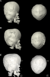
( A ) Preoperative three-dimensional (3D) computed tomography (CT) of the skull in a patient with Apert's syndrome with brachyturricephaly due to bilateral coronal and lambdoid synostosis. ( B ) Postoperative 3D CT of the skull following posterior vault distraction osteogenesis showing improved brachycephaly. ( C ) Postoperative 3D CT of the skull following mid-vault distraction osteogenesis.
Fig. 4
Fig. 4
Fronto-orbital distraction osteogenesis with schematic showing placement of fronto-orbital osteotomies.
Fig. 5
Fig. 5
Pre- and postoperative three-dimensional (3D) computed tomography (CT) of the skull of a patient undergoing monobloc distraction osteogenesis.
Fig. 6
Fig. 6
Le Fort I, II, and III osteotomies utilized for distraction osteogenesis.
Fig. 7
Fig. 7
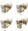
Kaban–Pruzansky classification of mandibular hypoplasia. Type I: The mandibular morphology is normal but small. Type IIA: Short ramus with adequate position of the glenoid fossa. Type IIB: Short ramus with abnormal inferior, medial, and anterior positioning of the temporomandibular joint with a hypoplastic condyle. Type III: Complete absence of the ramus, glenoid fossa, and temporomandibular joint.
Fig. 8
Fig. 8
( A ) Schematic of inverted- L osteotomy for vertical lengthening of the ramus in mandibular distraction osteogenesis. ( B ) Vertical symphyseal osteotomy for mandibular symphyseal distraction osteogenesis for correction of transverse deficiencies.
Fig. 9
Fig. 9
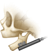
Internal distractor placement used in neonates with Pierre Robin sequence.
Fig. 10
Fig. 10
( A ) Preoperative pictures of a patient with right mandibular micrognathia. ( B ) Status post mandibular distraction osteogenesis.

References

    1. Cherkashin A. Distraction osteogenesis: origins and evolution. Distraction Osteogenesis Tissue Eng. 1998;34:1–35.
    1. Al-Namnam N MN, Hariri F, Rahman Z AA. Distraction osteogenesis in the surgical management of syndromic craniosynostosis: a comprehensive review of published papers. Br J Oral Maxillofac Surg. 2018;56(05):353–366. - PubMed
    1. Garrigues G E, Ledford C, Fitch R D. Ilizarov: the man, the myth, the method. An orthopaedic inspiration. Duke Orthop J. 2013;3(01):104–107.
    1. Hopper R A, Ettinger R E, Purnell C A, Dover M S, Pereira A R, Tunçbilek G. Thirty years later: what has craniofacial distraction osteogenesis surgery replaced? Plast Reconstr Surg. 2020;145(06):1073e–1088e. - PubMed
    1. Mundinger G S, Rehim S A, Johnson O, III et al.Distraction osteogenesis for surgical treatment of craniosynostosis: a systematic review. Plast Reconstr Surg. 2016;138(03):657–669. - PubMed