. 2024 Apr 9;149(15):1183-1201.
doi: 10.1161/CIRCULATIONAHA.123.064735. Epub 2023 Dec 15.

HEG1 Protects Against Atherosclerosis by Regulating Stable Flow-Induced KLF2/4 Expression in Endothelial Cells

Affiliations

HEG1 Protects Against Atherosclerosis by Regulating Stable Flow-Induced KLF2/4 Expression in Endothelial Cells

Ian A Tamargo et al. Circulation. .

Abstract

Background: Atherosclerosis preferentially occurs in arterial regions of disturbed blood flow, and stable flow (s-flow) protects against atherosclerosis by incompletely understood mechanisms.

Methods: Our single-cell RNA-sequencing data using the mouse partial carotid ligation model was reanalyzed, which identified Heart-of-glass 1 (HEG1) as an s-flow-induced gene. HEG1 expression was studied by immunostaining, quantitive polymerase chain reaction, hybridization chain reaction, and Western blot in mouse arteries, human aortic endothelial cells (HAECs), and human coronary arteries. A small interfering RNA-mediated knockdown of HEG1 was used to study its function and signaling mechanisms in HAECs under various flow conditions using a cone-and-plate shear device. We generated endothelial-targeted, tamoxifen-inducible HEG1 knockout (HEG1iECKO) mice. To determine the role of HEG1 in atherosclerosis, HEG1iECKO and littermate-control mice were injected with an adeno-associated virus-PCSK9 [proprotein convertase subtilisin/kexin type 9] and fed a Western diet to induce hypercholesterolemia either for 2 weeks with partial carotid ligation or 2 months without the surgery.

Results: S-flow induced HEG1 expression at the mRNA and protein levels in vivo and in vitro. S-flow stimulated HEG1 protein translocation to the downstream side of HAECs and release into the media, followed by increased messenger RNA and protein expression. HEG1 knockdown prevented s-flow-induced endothelial responses, including monocyte adhesion, permeability, and migration. Mechanistically, HEG1 knockdown prevented s-flow-induced KLF2/4 (Kruppel-like factor 2/4) expression by regulating its intracellular binding partner KRIT1 (Krev interaction trapped protein 1) and the MEKK3-MEK5-ERK5-MEF2 pathway in HAECs. Compared with littermate controls, HEG1iECKO mice exposed to hypercholesterolemia for 2 weeks and partial carotid ligation developed advanced atherosclerotic plaques, featuring increased necrotic core area, thin-capped fibroatheroma, inflammation, and intraplaque hemorrhage. In a conventional Western diet model for 2 months, HEG1iECKO mice also showed an exacerbated atherosclerosis development in the arterial tree in both sexes and the aortic sinus in males but not in females. Moreover, endothelial HEG1 expression was reduced in human coronary arteries with advanced atherosclerotic plaques.

Conclusions: Our findings indicate that HEG1 is a novel mediator of atheroprotective endothelial responses to flow and a potential therapeutic target.

Keywords: KLF2/4; atherosclerosis; endothelial cells; heart-of-glass 1; mechanobiology.


Conflict of interest statement

Disclosures Dr Jo is the founder of Flokines Pharma. The other authors report no conflicts.

Figures

Fig. 1.
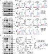
Fig. 1.. Heg1 expression is regulated by flow in mouse artery endothelial cells (ECs) in vivo.
(a) scRNA-seq data of the mouse right (RCAs) exposed to stable flow (s-flow) and left carotid arteries (LCAs) exposed to disturbed flow (d-flow) obtained at 2 days or 2 weeks post-partial carotid ligation (PCL) surgery were analyzed. GC (greater curvature) and LC (lesser curvature) in the aortic arch are shown. Heg1 expression is increased in ECs exposed to s-flow (E1–4) in the RCA compared to the d-flow exposed LCA ECs (E5–8). (b-d) Flow-sensitive Heg1 mRNA expression in mouse carotid ECs was verified by (b) gene array analysis of 1 day to 2-week post-PCL datasets (n=5), (c) qPCR at 2 weeks post-PCL (n=7), and (d) hybridization chain reaction (HCR) mRNA imaging at 2 weeks post-PCL (n=3). Gene array, qPCR, and HCR quantifications are presented as mean fold change ± SEM. P-values were calculated by two-tailed unpaired Student’s t-test, except for the 2-week gene array data in (b), which was analyzed by a two-tailed unpaired Mann-Whitney U-test due to non-normal distribution.
Fig. 2.
Fig. 2.. Unidirectional laminar shear stress (ULS) induces HEG1 mRNA and protein expression, stimulates protein secretion into the media and translocation toward downstream side in human aortic ECs (HAECs) in vitro.
(a-i) HAECs were exposed to static (no-flow), ULS (simulating s-flow), OSS (oscillatory shear stress simulating d-flow) conditions for various time-dependent studies: 24 hr (a-c, j), 3 to 24 hr time course (d-f), 30 min to 3 hr time course (g-i), or 2 to 30 min time course (k). Cell lysates or conditioned media (CM) were analyzed by western blots using calnexin antibody or Coomassie staining of bovine serum albumin (BSA) as controls (a, c, d, f, g, i) and qPCR using 18S as a control (b, e, h). HEG1 mRNA and protein increase in the lysate in a time-dependent manner by ULS (a, b, d, e). HEG1 protein is secreted to the CM by ULS in a time-dependent manner (c, f, i). During the first 3 hr of ULS, HEG1 protein in the lysate is reduced while increasing in the media at the same time, without changing its mRNA levels (g-i), showing that HEG1 is secreted. (j, k) Immunostaining of HAECs using the antibodies to HEG1 and VE-cadherin (VE-Cad) shows that HEG1 protein is randomly distributed in ECs in 24 hr static and OSS conditions but accumulates in the downstream side in ECs exposed to 24 hr ULS. Time course study shows that ULS rapidly induces translocation of randomly distributed HEG1 to the downstream side near cell-cell junctions as early as 2 min. n=6–7 for (a), n=7–11 for (b), n=3 for (c), n=3–7 for (e), n=3–5 for (f), n=6 for (h), n=3 for (i), n=35–76 for (j), and n=19–21 for (k). Western blot and qPCR quantifications presented as mean fold change ± SEM. P-values were calculated by ordinary one-way ANOVA for (a-i), Kruskal-Wallis one-way ANOVA for mRNA data in (e) and (h) due to non-normal distribution, and mixed effects two-way ANOVA for (j, k).
Fig. 3.
Fig. 3.. HEG1 mediates ULS-induced protective responses of HAECs.
HAECs were treated with HEG1 siRNA (siHEG1) or control siRNA (siCtrl) for 24 hr, followed by ULS for 1 day in cone and plate viscometer (a, c) or 3 days in IBIDI pump system (b). Following shear, THP-1 monocyte adhesion (n=6) (a), FITC-avidin permeability (n=3–12) (b), and endothelial scratch wound healing (n=6) (c) assays were performed, showing that HEG1 knockdown prevents ULS-mediated protective effects in HAECs. (d, e) Western blots show that HEG1 knockdown prevents ULS-induced suppression of VCAM1 expression (n=8) (d), and stimulation of eNOS expression (n=7–11) (e). Monocyte adhesion was quantified as mean number of fluorescent THP-1 monocytes per field ± SEM. Migration quantified as mean percent reduction in 0 hr scratch wound area ± SEM. All other quantifications presented as mean fold change ± SEM. P-values were all calculated using ordinary one-way ANOVA, apart from monocyte adhesion in (a), and HEG1 protein in (d), which were calculated with Kruskal-Wallis one-way ANOVA due to non-normality.
Fig. 4.
Fig. 4.. HEG1 is a critical mediator of ULS-induced KLF2/4 expression in HAECs by the MEKK3-MEK5-ERK5 pathway.
(a-c) HAECs treated with siHEG1 or siCtrl for 1 day were exposed to static or ULS for 3 hr to determine the signaling pathways mediating KLF2/4 expression by western blot (n=6–8) and qPCR (n=6–8). HEG1 knockdown inhibits ULS-induced ERK5 phosphorylation and KLF2/4 mRNA and protein expression. (d-f) Treatment of HAECs with MEKK3 inhibitor Ponatinib (Pon, 2 μM) prevents ULS-induced ERK5 phosphorylation and KLF2/4 mRNA and protein expression without affecting HEG1 expression (n=4–5). (g, h) HAECs were co-treated with siHEG1 or siCtrl, and adeno-KLF4 (Ad-KLF4) or adeno-GFP particles (Ad-GFP) for 1 day, followed by 24 hr ULS or static conditions. KLF4 overexpression restores the effects of ULS on the expression of eNOS and VCAM1 (n=4–5), and migration (n=3) in HEG1 knockdown ECs. Migration quantified as mean percent reduction in 0 hr scratch wound area ± SEM. All other quantifications presented as mean fold change ± SEM. P-values were calculated using two-way ANOVA with Tukey’s post-hoc test for (b, c, e, f) and ordinary one-way ANOVA for (g, h).
Fig. 5.
Fig. 5.. ULS stimulates the ERK5-KLF2/4 pathway by reducing intracellular KRIT1 protein levels through release in a HEG1-dependent manner.
(a) HAECs treated with siKRIT1 or siCtrl for 1 day were exposed to static or ULS for 3 hr, and the lysates were analyzed by western blot and qPCR. KRIT1 knockdown increases ERK5 phosphorylation and KLF2/4 mRNA and protein expression (n=4). (b) Western blot analysis of HAECs exposed to static, ULS, or OSS conditions for 24 hr showed that KRIT1 protein levels were reduced, while KRIT1 mRNA level increased in the lysates of cells exposed to ULS as compared to static or OSS (n=6–8). (c) In the same study, KRIT1 protein was found only in the conditioned media (CM) of HAECs exposed to ULS, but not in static or OSS conditions (n=3). (d) HEG1 siRNA knockdown blocks 24 hr ULS-induced KRIT1 release into the conditioned media in HAECs (n=3). (e) HEG1 knockdown inhibits the reduction of KRIT1 protein in the cell lysate by 3 hr ULS in HAECs (n=12–13). (f) HEG1 and KRIT1 remain bound to each other in both static and ULS conditions. MEKK3 is not pulled down along with HEG1 and KRIT1 in HAECs (n=3). Quantifications presented as mean fold change ± SEM. P-values were calculated using ordinary one-way ANOVA (a, b, c, e), and two-tailed unpaired Student’s t-test (d, f).
Fig. 6.
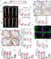
Fig. 6.. EC-specific HEG1 knockout in mice (HEG1iECKO) stimulates development of advanced atherosclerotic plaques in the PCL model.
(a) Heg1 HCR mRNA imaging and VE-Cadherin protein immunofluorescent imaging of en face abdominal aorta shows EC knockout in HEG1iECKO mice compared to the littermate controls (HEG1WT) (n=17–19). (b) qPCR analysis of endothelial-enriched RNA samples of thoracic aorta shows Heg1 knockout and significant decreases in Klf2, Klf4, and Nos3 mRNA expression in HEG1iECKO mice. qPCR quantifications are presented as transcripts per million 18S control (n=13–23). (c) Male and female HEG1iECKO and HEG1WT mice treated with tamoxifen, AAV-PCSK9, and Western diet received PCL surgery on their LCAs. (d) Gross LCA image analysis shows increased atherosclerosis in HEG1iECKO mice at 2 weeks post-PCL (n=22–26). Longitudinal sections of the upper LCA (brackets) stained with Oil red O (ORO) (e) and Movat’s pentachrome (f) show increased lipid lesion area (g), necrotic core area (h), intima-media thickness (IMT) (i), and fibrinoid/muscle tissue staining (j) in HEG1iECKO mice compared to HEG1WT (n=21–26). (k) HEG1iECKO mice have increased CD68 positive leukocyte infiltration into LCA wall (n=10). (l) HEG1WT and HEG1iECKO mice had equivalent plasma concentrations of total-, LDL-, and HDL-cholesterol, as well as triglycerides (n=21–27). All quantifications presented as mean ± SEM. P-values were calculated using two-tailed unpaired Student’s t-test for (a, d, g, i, k, l) and two-tailed unpaired Mann-Whitney U-test for (b, h, j) due to non-normality.
Fig. 7.
Fig. 7.. HEG1iECKO mice develop increased atherosclerotic plaque in 2-month, chronic atherosclerosis model in a sex- and location-dependent manner.
(a) HEG1iECKO and HEG1WT mice treated with tamoxifen were injected with AAV-PCSK9 and fed a Western diet for 8 weeks. (b) En face ORO staining of the arterial tree shows increased lesion development in the aortic arch in male and female and the descending aorta of male HEG1iECKO mice compared to HEG1WT. (n=19–30). (c) ORO staining of the aortic sinus showed increased lesion development in male HEG1iECKO compared to the WT mice (n=15–26). (d, e) Movat’s pentachrome (n=18–31) (d) and CD68 (n=16–19) (e) staining of the aortic sinus reveal no major differences between HEG1iECKO and WT mice, except for a decrease in collagen staining in male HEG1iECKO. (f) HEG1iECKO and WT mice show equivalent plasma concentrations of total-, LDL-, and HDL-cholesterol, as well as triglycerides, within the same sex (n=18–32). Quantifications presented as mean ± SEM. P-values were calculated using two-tailed unpaired Student’s t-test.
Fig. 8.
Fig. 8.. HEG1 expression is significantly reduced in human coronary arteries with advanced plaques.
(a-b) Human coronary arteries containing varying degrees of atherosclerotic lesions were stained with HEG1 antibody (red) and DAPI (blue). (a) Shown are Hematoxylin and eosin staining images representing varying degrees of atherosclerotic lesions using American Heart Association plaque severity grades. (b) HEG1 fluorescence intensity in the endothelial layer was reduced in grade III-IV and V-VI plaques as compared to grade I-II plaques. Data are from 55 different patients and 180 sections (n=45–67 images per group). Quantifications presented as mean fluorescence intensity ± SEM. P-values were calculated using Kruskal-Wallis one-way ANOVA due to non-normality. (c) Proposed mechanism of HEG1 regulation and action in response to d-flow (OSS) and s-flow (ULS) in ECs. Under d-flow conditions, HEG1 protein is expressed at low levels, randomly located in the EC membrane, and bound to KRIT1, which keeps MEKK3 inactive, leading to low KLF2/4 expression and pro-atherogenic phenotype. In response to s-flow, HEG1 protein translocates to the downstream side and is secreted to the media with KRIT1, causing activation of the MEKK3-MEK5-ERK5-MEF2 pathway that induces KLF2/4 expression. KLF2/4, a well-known S-flow-induced transcription factor, then induces the expression of hundreds of target genes that lead to the maintenance of EC homeostasis and atheroprotection. Schematic created with BioRender.com.

Comment in

References

    1. Caro CG, Fitz-Gerald JM and Schroter RC. Arterial wall shear and distribution of early atheroma in man. Nature. 1969;223:1159–60. - PubMed
    1. Malek AM, Alper SL and Izumo S. Hemodynamic shear stress and its role in atherosclerosis. JAMA. 1999;282:2035–42. - PubMed
    1. VanderLaan PA, Reardon CA and Getz GS. Site specificity of atherosclerosis: site-selective responses to atherosclerotic modulators. Arterioscler Thromb Vasc Biol. 2004;24:12–22. - PubMed
    1. Li YS, Haga JH and Chien S. Molecular basis of the effects of shear stress on vascular endothelial cells. J Biomech. 2005;38:1949–71. - PubMed
    1. Demos C, Tamargo I and Jo H. Biomechanical regulation of endothelial function in atherosclerosis. In: Ohayon J, Finet G and Pettigrew R, eds. Biomechanics of Coronary Atherosclerotic Plaque: Academic Press; 2021(4): 3–47.

Publication types