. 2024 Feb 1;326(2):H418-H425.
doi: 10.1152/ajpheart.00729.2023. Epub 2023 Dec 15.

Empagliflozin treatment rescues abnormally reduced Na+ currents in ventricular cardiomyocytes from dystrophin-deficient mdx mice

Affiliations

Empagliflozin treatment rescues abnormally reduced Na+ currents in ventricular cardiomyocytes from dystrophin-deficient mdx mice

Jakob Sauer et al. Am J Physiol Heart Circ Physiol. .

Abstract

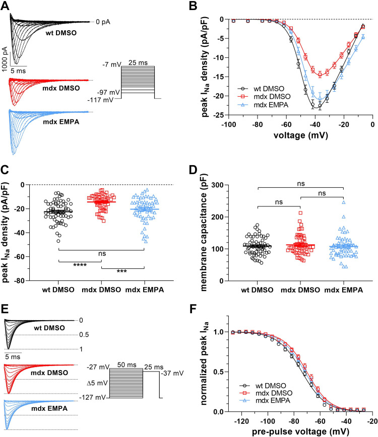
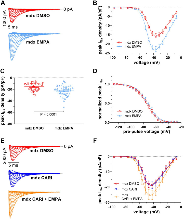
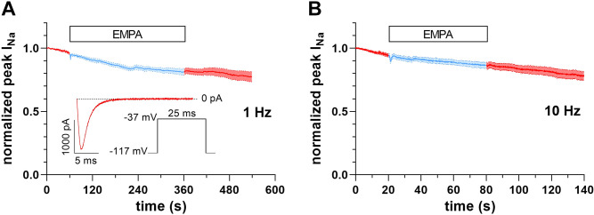
Cardiac arrhythmias significantly contribute to mortality in Duchenne muscular dystrophy (DMD), a severe muscle illness caused by mutations in the gene encoding for the intracellular protein dystrophin. A major source for arrhythmia vulnerability in patients with DMD is impaired ventricular impulse conduction, which predisposes for ventricular asynchrony, decreased cardiac output, and the development of reentrant circuits. Using the dystrophin-deficient mdx mouse model for human DMD, we previously reported that the lack of dystrophin causes a significant loss of peak Na+ current (INa) in ventricular cardiomyocytes. This finding provided a mechanistic explanation for ventricular conduction defects and concomitant arrhythmias in the dystrophic heart. In the present study, we explored the hypothesis that empagliflozin (EMPA), an inhibitor of sodium/glucose cotransporter 2 in clinical use to treat type II diabetes and nondiabetic heart failure, rescues peak INa loss in dystrophin-deficient ventricular cardiomyocytes. We found that INa of cardiomyocytes derived from mdx mice, which had received clinically relevant doses of EMPA for 4 wk, was restored to wild-type level. Moreover, incubation of isolated mdx ventricular cardiomyocytes with 1 µM EMPA for 24 h significantly increased their peak INa. This effect was independent of Na+-H+ exchanger 1 inhibition by the drug. Our findings imply that EMPA treatment can rescue abnormally reduced peak INa of dystrophin-deficient ventricular cardiomyocytes. Long-term EMPA administration may diminish arrhythmia vulnerability in patients with DMD.NEW & NOTEWORTHY Dystrophin deficiency in cardiomyocytes leads to abnormally reduced Na+ currents. These can be rescued by long-term empagliflozin treatment.

Keywords: Duchenne muscular dystrophy; arrhythmias; cardiomyocyte sodium currents; empagliflozin; mdx mice.


Conflict of interest statement

No conflicts of interest, financial or otherwise, are declared by the authors.

Figures

Figure 1.
Figure 1.
Na+ current (INa) properties in ventricular cardiomyocytes derived from wild-type (WT DMSO), untreated control mdx (mdx DMSO) and EMPA-treated mdx (mdx EMPA) mice. The latter mdx mouse cohort had received 15 mg/kg/day EMPA via the drinking water for 4 wk; WT and control mdx mice had received a respective concentration of DMSO. A: typical original current traces of a WT, a control mdx, and an EMPA-treated mdx cardiomyocyte, elicited by the pulse protocol displayed (inset). B: from a series of such experiments [n = 61 cells from 4 WT hearts (black); n = 56 cells from 4 control mdx hearts (red); n = 58 cells from 4 mdx EMPA hearts (light blue)], current density-voltage relationships were derived. Data points are represented as means ± SE. Parameters for INa activation derived from fits of the current-voltage relationships (function described in methods) are given in Table 1. C: dot plot comparing the maximum peak INa densities of WT, control mdx, and EMPA-treated mdx cardiomyocytes at −37 mV. ****P < 0.0001, significant difference between WT and mdx. ***P < 0.001, significant difference between mdx and mdx EMPA. ns, not significant (P = 0.19). D: dot plot comparing membrane capacitance values for cell size estimation of WT, control mdx, and EMPA-treated mdx cardiomyocytes. ns, not significant (P always > 0.38). E: original INa traces of a WT, a control mdx, and an EMPA-treated mdx cardiomyocyte, elicited by a 25-ms test pulse following a series of inactivating 50-ms prepulses. The pulse protocol used to test the voltage dependence of steady-state fast inactivation is displayed (inset). F: voltage dependencies of steady-state inactivation in WT, control mdx, and mdx EMPA cardiomyocytes (n = 48 cells from 4 WT hearts; n = 31 cells from 4 control mdx hearts; n = 32 cells from 4 mdx EMPA hearts). Parameters for steady-state fast inactivation derived from fits with a Boltzmann function (see methods) are given in Table 1.
Figure 2.
Figure 2.
Effect of 24-h incubation with 1 µM empagliflozin (EMPA) on Na+ current (INa) properties of ventricular cardiomyocytes derived from mdx mice. A: typical original current traces of a control mdx (mdx DMSO) and an EMPA-treated (mdx EMPA) cardiomyocyte (for pulse protocol see Fig. 1A). B: from a series of such experiments [n = 36 mdx cells (red); n = 43 mdx EMPA cells (light blue); all cells originating from 4 mdx hearts], current density-voltage relationships were derived. C: dot plot comparing the maximum peak INa densities of control mdx and EMPA-treated mdx cardiomyocytes at −37 mV. P = 0.0001, significant difference between mdx and mdx EMPA. D: voltage dependencies of steady-state inactivation (for pulse protocol, see Fig. 1E) in control mdx and mdx EMPA cardiomyocytes (n = 15 mdx cells; n = 18 mdx EMPA cells; all cells originating from 4 mdx hearts). INa activation and steady-state fast inactivation parameters are given in Table 1. E: original current traces of a control mdx (mdx DMSO), a cariporide-treated mdx (mdx CARI, 24-h incubation at 10 µM concentration), and a cariporide-plus EMPA-treated (mdx CARI + EMPA) cardiomyocyte. F: current density-voltage relationships derived from a series of such experiments [n = 16 mdx cells (red); n = 19 mdx CARI cells (dark blue); n = 15 mdx CARI + EMPA cells (orange); all cells originating from 2 mdx hearts].
Figure 3.
Figure 3.
Lack of acute empagliflozin (EMPA) effects on peak Na+ current (INa) of mdx ventricular cardiomyocytes. A: peak INa before, during superfusion with bath solution containing 1 µM EMPA (light blue), and after washout over time (n = 13 cells from 2 mdx hearts). Data are expressed as means ± SE. Inset: pulse protocol, applied at 1-Hz frequency and elicited INa at the very beginning of the experiment. B: equivalent experiment as described in A at 10-Hz frequency (n = 15 cells from 2 mdx hearts).

References

    1. Kamdar F, Garry DJ. Dystrophin-deficient cardiomyopathy. J Am Coll Cardiol 67: 2533–2546, 2016. doi:10.1016/j.jacc.2016.02.081. - DOI - PubMed
    1. Duan D, Goemans N, Takeda S, Mercuri E, Aartsma-Rus A. Duchenne muscular dystrophy. Nat Rev Dis Primers 7: 13, 2021. doi:10.1038/s41572-021-00248-3. - DOI - PMC - PubMed
    1. Bulfield G, Siller WG, Wight PA, Moore KJ. X chromosome-linked muscular dystrophy (mdx) in the mouse. Proc Natl Acad Sci USA 81: 1189–1192, 1984. doi:10.1073/pnas.81.4.1189. - DOI - PMC - PubMed
    1. Gavillet B, Rougier JS, Domenighetti AA, Behar R, Boixel C, Ruchat P, Lehr HA, Pedrazzini T, Abriel H. Cardiac sodium channel Nav1.5 is regulated by a multiprotein complex composed of syntrophins and dystrophin. Circ Res 99: 407–414, 2006. doi:10.1161/01.RES.0000237466.13252.5e. - DOI - PubMed
    1. Koenig X, Dysek S, Kimbacher S, Mike AK, Cervenka R, Lukacs P, Nagl K, Dang XB, Todt H, Bittner RE, Hilber K. Voltage-gated ion channel dysfunction precedes cardiomyopathy development in the dystrophic heart. PLoS One 6: e20300, 2011. doi:10.1371/journal.pone.0020300. - DOI - PMC - PubMed

Publication types

LinkOut - more resources