. 2024 Feb;43(2):355-371.
doi: 10.1007/s10096-023-04721-7. Epub 2023 Dec 15.

Genotyping of European Toxoplasma gondii strains by a new high-resolution next-generation sequencing-based method

Affiliations

Genotyping of European Toxoplasma gondii strains by a new high-resolution next-generation sequencing-based method

M Joeres et al. Eur J Clin Microbiol Infect Dis. 2024 Feb.

Abstract

Purpose: A new high-resolution next-generation sequencing (NGS)-based method was established to type closely related European type II Toxoplasma gondii strains.

Methods: T. gondii field isolates were collected from different parts of Europe and assessed by whole genome sequencing (WGS). In comparison to ME49 (a type II reference strain), highly polymorphic regions (HPRs) were identified, showing a considerable number of single nucleotide polymorphisms (SNPs). After confirmation by Sanger sequencing, 18 HPRs were used to design a primer panel for multiplex PCR to establish a multilocus Ion AmpliSeq typing method. Toxoplasma gondii isolates and T. gondii present in clinical samples were typed with the new method. The sensitivity of the method was tested with serially diluted reference DNA samples.

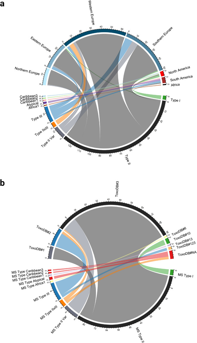
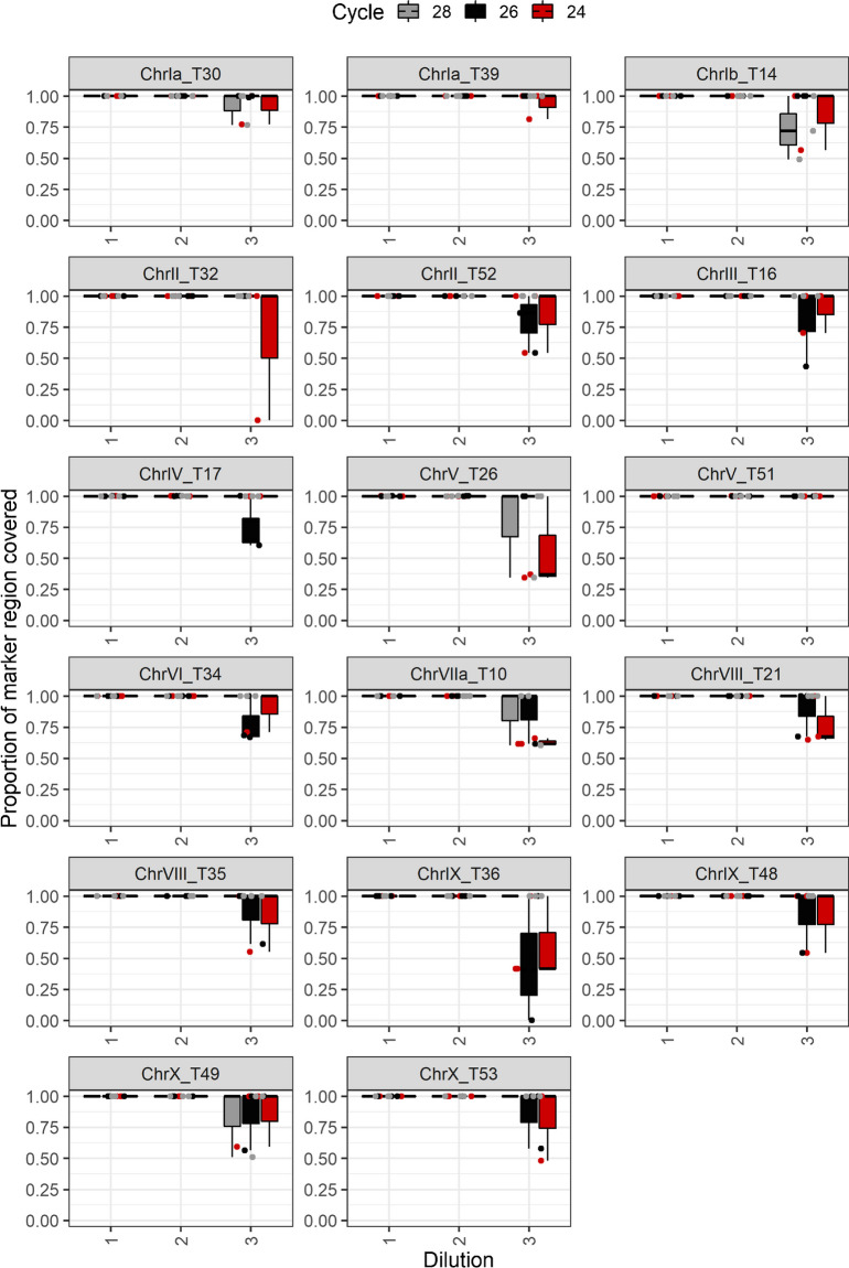
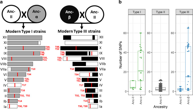
Results: Among type II specimens, the method could differentiate the same number of haplotypes as the reference standard, microsatellite (MS) typing. Passages of the same isolates and specimens originating from abortion outbreaks were identified as identical. In addition, seven different genotypes, two atypical and two recombinant specimens were clearly distinguished from each other by the method. Furthermore, almost all SNPs detected by the Ion AmpliSeq method corresponded to those expected based on WGS. By testing serially diluted DNA samples, the method exhibited a similar analytical sensitivity as MS typing.

Conclusion: The new method can distinguish different T. gondii genotypes and detect intra-genotype variability among European type II T. gondii strains. Furthermore, with WGS data additional target regions can be added to the method to potentially increase typing resolution.

Keywords: Discriminatory power; Highly polymorphic regions; Intra-genotype variability; Multilocus sequence typing; Toxoplasmosis; Typing.


Conflict of interest statement

The authors declare no competing interests.

Figures

Fig. 1
Fig. 1
Workflow of the establishment of the Ion AmpliSeq method. Created with BioRender.com
Fig. 2
Fig. 2
Geographic origin of 110 European T. gondii isolates and 47 clinical samples, which were genotyped with the Ion AmpliSeq method. The geographic origin of the reference strains PRU and CZ-H3 not genotyped with the Ion AmpliSeq method is also included. Thirteen non-European isolates, also genotyped with the Ion AmpliSeq method are excluded. Details are described in Supplementary Table 1
Fig. 3
Fig. 3
Microsatellite (MS) typing of T. gondii specimens using 15 markers. a Ten different categories of MS genotypes were reported for DNA from 170 specimens genotyped in this study and additionally for MS genotypes of the reference strains PRU and CZ-H3 described in the literature in relation to their regional origin. Seven regions were defined, consisting of Northern Europe (Denmark, Finland, Sweden, Norway), Eastern Europe (Austria, Czech Republic, Poland, Romania, Serbia, Slovakia), Southern Europe (Greece, Italy, Portugal, Spain), Western Europe (France, Germany, Netherlands, Switzerland, UK), Africa, North America, and South America. b Ten different categories of MS genotypes were reported for 123 isolates genotyped in this study and MS genotypes of the reference strains PRU and CZ-H3 described in the literature in relation to their PCR-RFLP genotyping results
Fig. 4
Fig. 4
SNP maps of all 14 T. gondii chromosomes based on the numbers of SNPs detected in non-overlapping windows of 333 bp in 43 type II genomes relative to the genome of ME49 (ToxoDB release 47). All identified highly polymorphic regions were categorized as first, second, third or fourth priority targets and their positions on the chromosomes are indicated with grey bars. Minimum and maximum numbers of SNPs per target are indicated on the right side of each chromosome. The 18 target regions used for Ion AmpliSeq typing are shown in orange
Fig. 5
Fig. 5
Coverage of 17 target regions by ME49 replicates used to assess the analytical sensitivity of the Ion AmpliSeq method after mapping to mapped the genome of ME49 (ToxoDB release 53). The proportion of coverage of each region is shown in relation to the different dilutions (1, 2, and 3 correspond to T. gondii DNA concentrations of 1 ng/μl, 0.1 ng/μl and 0.01 ng/ μl) and the number of PCR cycles
Fig. 6
Fig. 6
Comparison of the numbers of SNPs detected by Ion AmpliSeq typing in T. gondii specimens relative to the AmpliSeq-ME49-Reference per region and per genotype. The numbers of specimens per genotype are not equally distributed. The figure includes results of 121 type II specimens, twelve type II variants, 14 type III (excluding C25 and C26, classified as ToxoDB #123 by PCR-RFLP typing) and four type I specimens. Only one specimen each was analyzed in case of Africa 1 and Caribbean 1, 2 and 3 and in addition, two atypical specimens and five type II × III recombinants were examined. If a boxplot is missing, the affected region was not covered by the reads of the respective genotype and no SNPs could be reported. This was the case for Africa 1 in target region T30 as well as for the atypical specimens in target region T14. In addition, target regions T21 and T35 were not covered by the reads of type I, Africa 1, Caribbean 1, Caribbean 2 and atypical specimens
Fig. 7
Fig. 7
Neighbour-net analysis of T. gondii specimens based on SNPs detected by Ion AmpliSeq typing relative to the AmpliSeq-ME49-Reference in 17 target regions (software SplitsTree4). a Analysis of 164 specimens belonging to different genotypes revealed seven groups. All type II specimens are located in group A, type III in group C and type I in group G. Four type II × III recombinant strains (coloured in orange) are in group B and one in group A. Group D is represented by the genotypes Caribbean 1, 2 and 3, group E by two atypical strains and one specimen typed as Africa 1 is located in group F. b Analysis of 131 European type II specimens distinguishing specimens from North, East, South and West Europe. No clear regional patterns can be observed. However, different passages from the same isolates were identified as identical (No. 7 and 24; No. 8 and 19; No. 32 and 78) as were specimens from abortion outbreaks (No. 42–45 and 131; No. 48 and 49). Furthermore, eight specimens (No. 46, 51, 52, 56, 59, 62, 79, 85), which showed the same variation in the MS marker W35, were identical or very similar
Fig. 8
Fig. 8
Proposed genealogy of the T. gondii lineages type I and III and chromosome segregation during the proposed crosses, modified from Boyle et al. (2006) [37], combined with the number of SNPs detected by Ion AmpliSeq typing. a Chromosome segregation during the two proposed crosses (ancestral type II (Anc-II) × ancestral α (Anc-α) and ancestral type II × ancestral β (Anc-β)) (details in Supplementary Figure 4a-n). On the left (for type I) and right (for type III), all 14 chromosomes are represented schematically with their proposed ancestry coloured in grey (α), black (β), or white (type II). The positions and the names of the 17 Ion AmpliSeq target regions on the chromosomes are denoted in red. b Number of SNPs detected by Ion AmpliSeq typing within types I, II and III specimens relative to the AmpliSeq-ME49-Reference (details in Supplementary Figure 5). In case of type I and type III, the regions and the associated SNPs were differentiated into ancestral type II (Anc-II) and ancestral α (Anc-α) and ancestral β (Anc-β). Large numbers of SNPs per region are only observed in Ion AmpliSeq targets located in parts of the genome, for which Ancestral α or β origin was proposed

References

    1. Schlüter D, Däubener W, Schares G, et al. Animals are key to human toxoplasmosis. Int J Med Microbiol. 2014;304:917–929. doi: 10.1016/j.ijmm.2014.09.002. - DOI - PubMed
    1. Robert-Gangneux F, Dardé M-L. Epidemiology of and diagnostic strategies for toxoplasmosis. Clin Microbiol Rev. 2012;25:264–296. doi: 10.1128/CMR.05013-11. - DOI - PMC - PubMed
    1. Dubey JP. Toxoplasmosis of animals and humans. Third. Boca Raton: CRC Press; 2022.
    1. Stelzer S, Basso W, Benavides Silván J, et al. Toxoplasma gondii infection and toxoplasmosis in farm animals: risk factors and economic impact. Food Waterborne Parasitol. 2019;15:e00037. doi: 10.1016/j.fawpar.2019.e00037. - DOI - PMC - PubMed
    1. Dámek F, Swart A, Waap H, et al. Systematic review and modelling of age-dependent prevalence of Toxoplasma gondii in livestock, wildlife and felids in Europe. Pathogens. 2023;12:97. doi: 10.3390/pathogens12010097. - DOI - PMC - PubMed