

Into the fold: advances in understanding aPKC membrane dynamics
- PMID: 38100320
- PMCID: PMC10754278
- DOI: 10.1042/BCJ20230390
Into the fold: advances in understanding aPKC membrane dynamics
Abstract
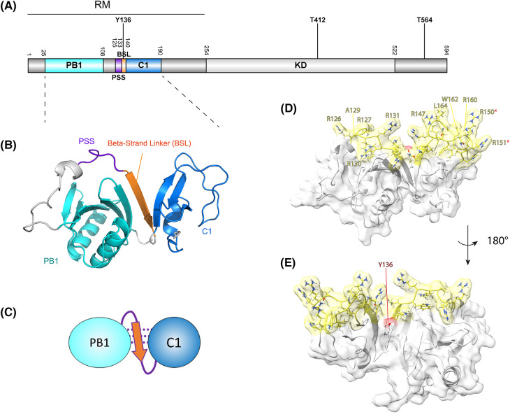
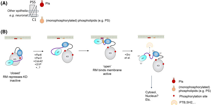
Atypical protein kinase Cs (aPKCs) are part of the PKC family of protein kinases and are atypical because they don't respond to the canonical PKC activators diacylglycerol (DAG) and Ca2+. They are central to the organization of polarized cells and are deregulated in several cancers. aPKC recruitment to the plasma membrane compartment is crucial to their encounter with substrates associated with polarizing functions. However, in contrast with other PKCs, the mechanism by which atypical PKCs are recruited there has remained elusive until recently. Here, we bring aPKC into the fold, summarizing recent reports on the direct recruitment of aPKC to membranes, providing insight into seemingly discrepant findings and integrating them with existing literature.
Keywords: apkc; atypical pkc; cell polarity; phospholipids; protein kinase c.
© 2023 The Author(s).
Conflict of interest statement
The authors declare that there are no competing interests associated with the manuscript.
Figures
Similar articles
-
Pleckstrin Homology (PH) Domain Leucine-rich Repeat Protein Phosphatase Controls Cell Polarity by Negatively Regulating the Activity of Atypical Protein Kinase C.J Biol Chem. 2016 Nov 25;291(48):25167-25178. doi: 10.1074/jbc.M116.740639. Epub 2016 Oct 19. J Biol Chem. 2016. PMID: 27760826 Free PMC article.
-
Possible role of atypical protein kinase C activated by arachidonic acid in Ca2+ sensitization of rabbit smooth muscle.J Physiol. 1997 Apr 1;500 ( Pt 1)(Pt 1):95-109. doi: 10.1113/jphysiol.1997.sp022002. J Physiol. 1997. PMID: 9097936 Free PMC article.
-
Autocrine Signaling Underlies Fast Repetitive Plasma Membrane Translocation of Conventional and Novel Protein Kinase C Isoforms in β Cells.J Biol Chem. 2016 Jul 15;291(29):14986-95. doi: 10.1074/jbc.M115.698456. Epub 2016 May 20. J Biol Chem. 2016. PMID: 27226533 Free PMC article.
-
How is protein kinase C activated in CNS.Neurochem Int. 1993 May;22(5):417-33. doi: 10.1016/0197-0186(93)90037-6. Neurochem Int. 1993. PMID: 8485448 Review.
-
Protein kinase C lambda/iota (PKClambda/iota): a PKC isotype essential for the development of multicellular organisms.J Biochem. 2003 Jan;133(1):9-16. doi: 10.1093/jb/mvg018. J Biochem. 2003. PMID: 12761193 Review.
References
Publication types
MeSH terms
Substances
Grants and funding
LinkOut - more resources
Full Text Sources
Miscellaneous