. 2023 Dec 15;9(50):eadj9974.
doi: 10.1126/sciadv.adj9974. Epub 2023 Dec 15.

Cryo-EM structure of influenza helical nucleocapsid reveals NP-NP and NP-RNA interactions as a model for the genome encapsidation

Affiliations

Cryo-EM structure of influenza helical nucleocapsid reveals NP-NP and NP-RNA interactions as a model for the genome encapsidation

Florian Chenavier et al. Sci Adv. .

Abstract

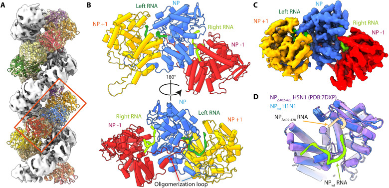
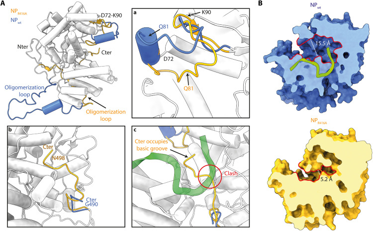
Influenza virus genome encapsidation is essential for the formation of a helical viral ribonucleoprotein (vRNP) complex composed of nucleoproteins (NP), the trimeric polymerase, and the viral genome. Although low-resolution vRNP structures are available, it remains unclear how the viral RNA is encapsidated and how NPs assemble into the helical filament specific of influenza vRNPs. In this study, we established a biological tool, the RNP-like particles assembled from recombinant influenza A virus NP and synthetic RNA, and we present the first subnanometric cryo-electron microscopy structure of the helical NP-RNA complex (8.7 to 5.3 Å). The helical RNP-like structure reveals a parallel double-stranded conformation, allowing the visualization of NP-NP and NP-RNA interactions. The RNA, located at the interface of neighboring NP protomers, interacts with conserved residues previously described as essential for the NP-RNA interaction. The NP undergoes conformational changes to enable RNA binding and helix formation. Together, our findings provide relevant insights for understanding the mechanism for influenza genome encapsidation.


Figures

Fig. 1.
Fig. 1.. Visualization of the right-handed double-stranded helical RNP-like by EM and AFM.
(A) EM observation of RNP-like sample by negative staining (sodium silicotungstate). Top, yellow circles highlight the presence of trimers, tetramers, and oligomeric rings of NP. The red boxes [(A) to (C)] correspond to the insets in the neighboring panels. (B) Vitrified RNP-like particles in cryo-conditions. (C) Observation of the RNP-like particles by AFM. Helical groves are indicated by green arrows. The black-orange-white scale indicates the selected z height of the sample, which corresponds to the topography of the particle in nanometers. White horizontal scale bars, 50 nm [(A) to (C)]. (D) Helically symmetrized cryo-EM 3D reconstruction of the RNP-like particle at 8.7 Å. The reconstruction displays about two intertwined helical turns. Each strand is colored with either a white-to-purple or a white-to-yellow color gradient. The positive sense of the helical strands is upward indicated by colored arrows.
Fig. 2.
Fig. 2.. Local reconstruction and NP atomic model.
(A) Fitting of symmetrized NP atomic model modified from PDB code: 2IQH. Each protomer is colored differently and displayed as cartoon representation. The boxed region corresponds to the area used for focused 3D reconstruction at 5.3-Å resolution. (B) Resulting pseudo-atomic model of three neighboring NP protomers, NP−1, NP, and NP+1 in red, blue, and yellow ribbons, respectively, and two RNA molecules at the NP-NP interface, RNA left and right in green and chartreuse ribbons, respectively. (C) Of note, the oligomerization loop of the NP+1 protomer is extrapolated on the basis of the oligomerization loop of protomers NP and NP−1 as it is not included in the focused reconstruction map. Top, view from the central axis of the helix of the atomic model build from the focused reconstruction map (5.3 Å). Bottom, view toward the center of the helix of the same nucleo-protomers. Oligomerization loops are clearly visible, entering the neighboring NP protomer. Focused 3D reconstruction map colored to corresponding NP protomers, NP−1, NP, and NP+1 in red, blue, and yellow, respectively. (D) Overlay of the central blue NP protomer with chartreuse left RNA, with the crystal structure of IAV H5N1 NP in purple, with three RNA nucleotides in beige (PDB code: 7DXP).
Fig. 3.
Fig. 3.. Visualization of the RNA molecules in regard to the NP central basic groove.
(A) Localization of the RNA molecules on the positively charged region of NP. Electrostatic surface representation of NP colored according to the residues charged positively (blue) or negatively (red). (B) Close-up views on the right and left RNAs shown in chartreuse and green ribbons; the NP residues most likely interacting with the RNA phosphate backbone, bases, or in the close vicinity of the RNA are shown respectively as sticks colored in orange, pink, and white, while the rest of the protein appears as blue ribbons.
Fig. 4.
Fig. 4.. Comparison of the NP conformations from the RNP-like helical structure (NPwt) with the monomeric NP R416A mutant crystal structure (NPR416A).
(A) Superimposition of the NP protomer from the RNP-like reconstruction (NPwt) and the monomeric NPR416A crystal structure (PDB code: 3ZDP). For clarity, RNA was omitted from NPwt. The global root mean square deviation (RMSD) is 0.83 Å, and the two structures are shown as a single chain colored in white, except for when the local RMSD is more than 3 Å; then, NPwt is displayed in blue and NPR416A in yellow. The oligomerization loop in NPR416A is folded inside the protein, while the oligomerization loop of NPwt appears pointing to the solvent. Box (a) corresponds to a close-up view on the D72-K90 loop, either folded in an α helix in NPwt (blue) or unfolded in NPR416A (yellow). Box (b) displays the C-terminal tail (G490-N498) either bound to the rest of the protein in NPR416A (yellow) or not visible in the NPwt EM density—Note that the RNA is omitted in this box. Box (c) shows the steric clash between the RNA molecule from NPwt and the C-terminal residues of NPR416A. (B) Central basic groove accessibility is modified by disordered D72-K90 loop folding. Surfaces of NPwt (top) and NPR416A (bottom) are presented in the same orientation and colored in blue and yellow, respectively, with the RNA molecule appearing in chartreuse cartoons. Surfaces are sliced up to the beginning of the NP-RNA central basic groove for clarity. Contour of the basic groove entrance is lined in red, and the size of the grove is indicated by the white dotted line.
Fig. 5.
Fig. 5.. Model of the full pathway of the RNA and schematic model of the vRNP.
(A) Proposed RNA pathway. Electrostatic surface representation of an NP protomer and two green ribbon RNA molecules as seen from the RNP-like focused reconstruction. The green dotted line corresponds to the proposed RNA pathway. (B) Surface representation of two neighboring NP protomers in dark gray and white, visualized from the center of the helix. The structure of the RNA from the cryo-EM structure appears in green ribbon representation, and the green dotted line corresponds to the proposed pathway of the rest of the RNA for each protomer. Boxed region, close up on one RNA molecule enclosed at the NP-NP interface visualized toward the center of the helix. (C) Schematic model of a vRNP (top corner); antiparallel helix modeled by symmetry from the RNP-like reconstruction; the RNA-dependent RNA polymerase is represented in sea green. NP protomers from one strand are colored as gradients from light to deep purple, and from light to dark yellow for the second strand. The RNA is colored in green; the arrow indicates a potential area for the formation of an extended loop of RNA.

Similar articles

Cited by

References

    1. D. McGeoch, P. Fellner, C. Newton, Influenza virus genome consists of eight distinct RNA species. Proc. Natl. Acad. Sci. U.S.A. 73, 3045–3049 (1976). - PMC - PubMed
    1. A. J. Eisfeld, G. Neumann, Y. Kawaoka, At the centre: Influenza A virus ribonucleoproteins. Nat. Rev. Microbiol. 13, 28–41 (2015). - PMC - PubMed
    1. S. R. Matzinger, T. D. Carroll, J. C. Dutra, Z.-M. Ma, C. J. Miller, Myxovirus resistance gene A (MxA) expression suppresses influenza A virus replication in alpha interferon-treated primate cells. J. Virol. 87, 1150–1158 (2013). - PMC - PubMed
    1. B. Mänz, D. Dornfeld, V. Götz, R. Zell, P. Zimmermann, O. Haller, G. Kochs, M. Schwemmle, Pandemic influenza a viruses escape from restriction by human mxa through adaptive mutations in the nucleoprotein. PLOS Pathog. 9, e1003279 (2013). - PMC - PubMed
    1. R. M. Pinto, S. Bakshi, S. Lytras, M. K. Zakaria, S. Swingler, J. C. Worrell, V. Herder, K. E. Hargrave, M. Varjak, N. Cameron-Ruiz, M. Collados Rodriguez, M. Varela, A. Wickenhagen, C. Loney, Y. Pei, J. Hughes, E. Valette, M. L. Turnbull, W. Furnon, Q. Gu, L. Orr, A. Taggart, O. Diebold, C. Davis, C. Boutell, F. Grey, E. Hutchinson, P. Digard, I. Monne, S. K. Wootton, M. K. L. MacLeod, S. J. Wilson, M. Palmarini, BTN3A3 evasion promotes the zoonotic potential of influenza A viruses. Nature 619, 338–347 (2023). - PubMed

Publication types