Case Reports
. 2023 Dec 14;11(12):e007463.
doi: 10.1136/jitc-2023-007463.

Immune microenvironment of basal cell carcinoma and tumor regression following combined PD-1/LAG-3 blockade

Affiliations
Case Reports

Immune microenvironment of basal cell carcinoma and tumor regression following combined PD-1/LAG-3 blockade

Julie Stein Deutsch et al. J Immunother Cancer. .

Abstract

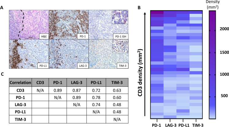
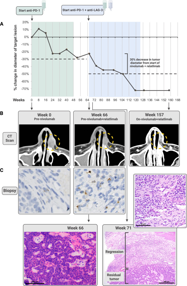
Systemic treatment options for patients with locally advanced or metastatic basal cell carcinoma (BCC) are limited, particularly when tumors are refractory to anti-programmed cell death protein-1 (PD-1). A better understanding of immune checkpoint expression within the BCC tumor microenvironment may inform combinatorial treatment strategies to optimize response rates. CD3, PD-1, programmed death ligand-1 (PD-L1), lymphocyte activation gene 3 (LAG-3), and T-cell immunoglobulin domain and mucin domain 3 (TIM-3)+ cell densities within the tumor microenvironment of 34 archival, histologically aggressive BCCs were assessed. Tumor infiltrating lymphocyte (TIL) expression of PD-1, PD-L1, and LAG-3, and to a lesser degree TIM-3, correlated with increasing CD3+ T-cell densities (Pearson's r=0.89, 0.72, 0.87, and 0.63, respectively). 100% of BCCs (34/34) demonstrated LAG-3 and PD-1 expression in >1% TIL; and the correlation between PD-1 and LAG-3 densities was high (Pearson's r=0.89). LAG-3 was expressed at ~50% of the level of PD-1. Additionally, we present a patient with locally-advanced BCC who experienced stable disease during and after 45 weeks of first-line anti-PD-1 (nivolumab), followed by a partial response after the addition of anti-LAG-3 (relatlimab). Longitudinal biopsies throughout the treatment course showed a graduated increase in LAG-3 expression after anti-PD-1 therapy, lending support for coordinated immunosuppression and suggesting LAG-3 as a co-target for combination therapy to augment the clinical impact of anti-PD-(L)1.

Keywords: biomarkers, tumor; carcinoma, basal cell; immunohistochemistry; pathology; tumor microenvironment.


Conflict of interest statement

Competing interests: JSD receives research funding from Bristol-Myers Squibb. DW is an employee of Merck. EJL serves as a consultant/advisory board member for Array BioPharma, Bristol-Myers Squibb, EMD Serono, Genentech, Macrogenics, Merck, Millennium, Novartis, Sanofi/Regeneron, and receives institutional research funding from Bristol-Myers Squibb, Merck and Regeneron. JMT serves as a consultant/advisory board member for Bristol-Myers Squibb, Merck, AstraZeneca, Compugen, and Akoya Biosciences. No additional potential conflicts of interest were disclosed.

Figures

Figure 1
Figure 1
Patterns of immunoactive marker expression in aggressive basal cell carcinomas (BCCs). (A) Photomicrograph (top left) displaying a representative tumor-immune cell interface within BCC (immune cells on left and tumor on right of black dotted line, H&E staining). Programmed cell death protein-1 (PD-1) immunohistochemistry highlights PD-1 expression by tumor infiltrating lymphocyte (TIL) and tumor cells (TCs) (top middle). PD-1 amplified in-situ hybridization (ISH) was used to verify the PD-1 expression by TCs (top right). Inset shows positive signal in the cytoplasm (red, punctate dots). Programmed death ligand-1 (PD-L1), lymphocyte activation gene 3 (LAG-3), and T-cell immunoglobulin domain and mucin domain 3 (TIM-3) expressions were also present on TIL in this same region. Scale bar=50 µm. (B) Heat map relating CD3+ T-cell densities with PD-1, PD-L1 LAG-3, and TIM-3 lymphocyte expression on a per-specimen basis. Specifically, each row in the heat map represents one specimen from an individual patient. In contrast to the other markers, TIM-3 densities were relatively low and did not associate with an increasing T-cell infiltrate. (C) Pearson’s correlation coefficients, r, showing the strong relationship between CD3, PD-1, and LAG-3 expression.
Figure 2
Figure 2
The patient whose advanced basal cell carcinoma stabilized on anti-programmed cell death protein-1 (PD-1), then regressed following the addition of anti-lymphocyte activation gene 3 (LAG-3) therapy. (A) the patient received nivolumab (anti-PD-1) over 48 weeks (green) during which he experienced stable disease per Response Evaluation Criteria in Solid Tumors (RECIST) V.1.1. The tumor remained stable in size for 18 weeks after discontinuation of therapy, at which point the patient received nivolumab plus relatlimab (anti-LAG-3; blue). He experienced a partial response 39 weeks later, ongoing at 21.5 months from nivolumab/relatlimab initiation. Dashed lines show a 30% decrease in tumor diameter from baseline during each treatment regimen. Both CT scans (B) and paired pretreatment and on-treatment biopsies stained with H&E (C) demonstrate tumor regression. The high-powered view of the regression area shown for Week 71 shows fibrosis, plasma cells, foamy macrophages and numerous lymphocytes. The LAG-3 immunohistochemistry (IHC) staining performed on the Week 0 (immediate pretreatment) and Week 66 biopsies shows an increase in LAG-3 expression after administration of anti-PD-1 monotherapy prior to anti-PD-1+LAG-3 treatment. The patient went on to demonstrate an objective response to combinatorial therapy. Additional images for LAG-3 expression over the course of therapy are shown in online supplemental figure S3. Some figure elements created with BioRender.com.

References

    1. Tay EY-X, Teoh Y-L, Yeo MS-W. Hedgehog pathway inhibitors and their utility in basal cell carcinoma: A comprehensive review of current evidence. Dermatol Ther (Heidelb) 2019;9:33–49. 10.1007/s13555-018-0277-7 - DOI - PMC - PubMed
    1. Stratigos AJ, Sekulic A, Peris K, et al. . Cemiplimab in locally advanced basal cell carcinoma after hedgehog inhibitor therapy: an open-label, multi-centre, single-arm, phase 2 trial. Lancet Oncol 2021;22:848–57. 10.1016/S1470-2045(21)00126-1 - DOI - PubMed
    1. Lipson EJ, Lilo MT, Ogurtsova A, et al. . Basal cell carcinoma: PD-L1/PD-1 Checkpoint expression and tumor regression after PD-1 blockade. J Immunother Cancer 2017;5:23. 10.1186/s40425-017-0228-3 - DOI - PMC - PubMed
    1. Lei Q, Wang D, Sun K, et al. . Resistance mechanisms of anti-Pd1/Pdl1 therapy in solid tumors. Front Cell Dev Biol 2020;8:672. 10.3389/fcell.2020.00672 - DOI - PMC - PubMed
    1. Duffield AS, Ascierto ML, Anders RA, et al. . Th17 immune Microenvironment in Epstein-Barr virus-negative Hodgkin lymphoma: implications for Immunotherapy. Blood Adv 2017;1:1324–34. 10.1182/bloodadvances.2017007260 - DOI - PMC - PubMed

Publication types

MeSH terms

Substances

LinkOut - more resources