Review
. 2024 Apr:160:130-152.
doi: 10.1016/j.clinph.2023.11.015. Epub 2023 Nov 30.

The blink reflex and its modulation - Part 1: Physiological mechanisms

Affiliations
Review

The blink reflex and its modulation - Part 1: Physiological mechanisms

Markus Kofler et al. Clin Neurophysiol. 2024 Apr.

Erratum in

Abstract

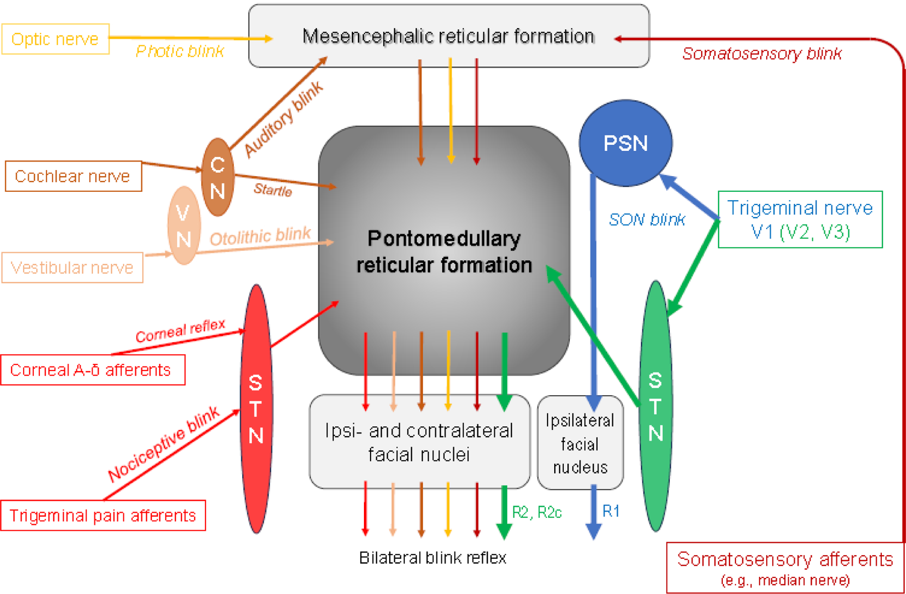
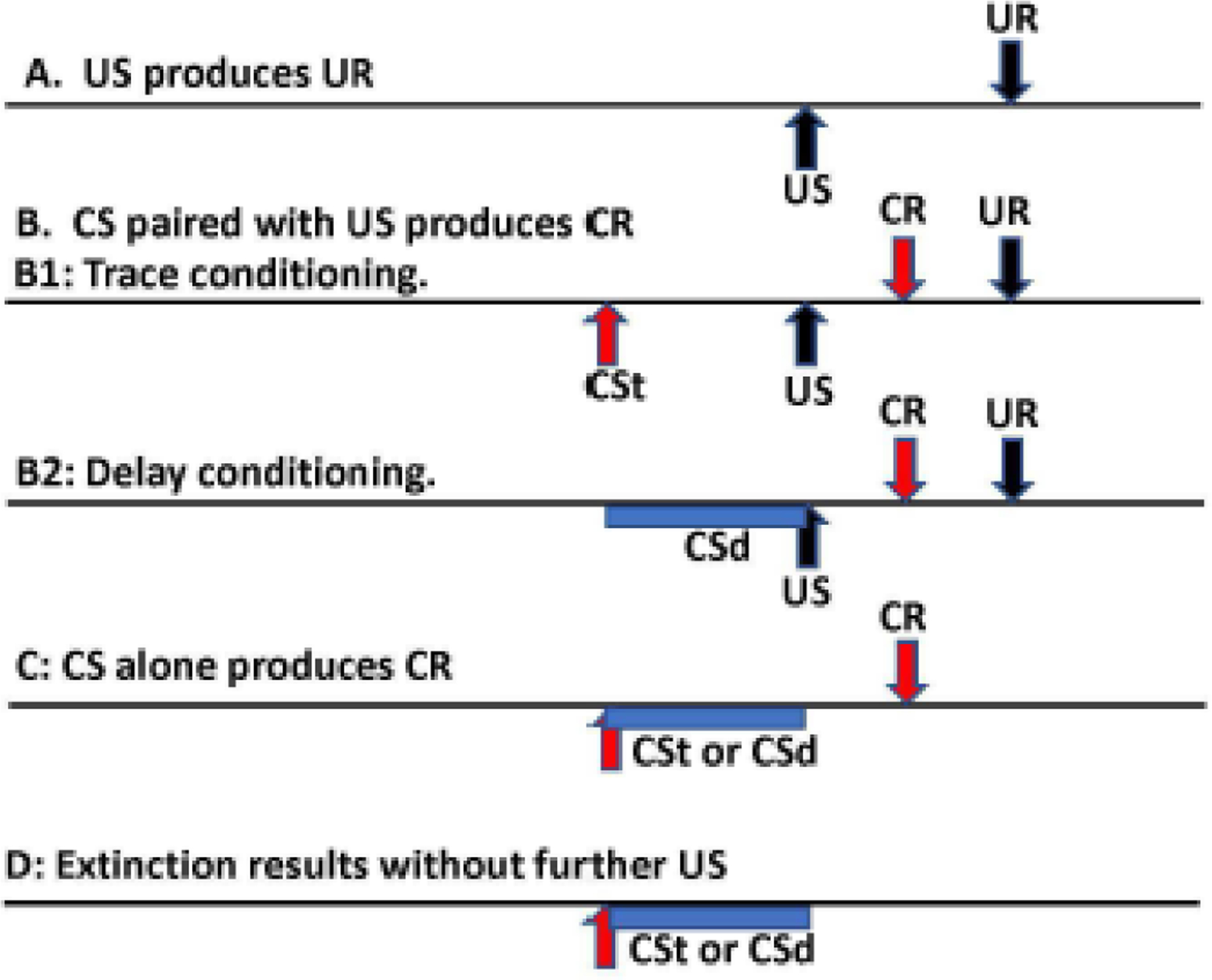
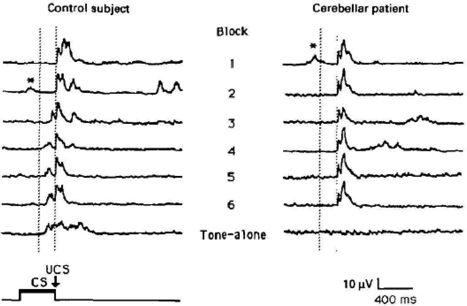
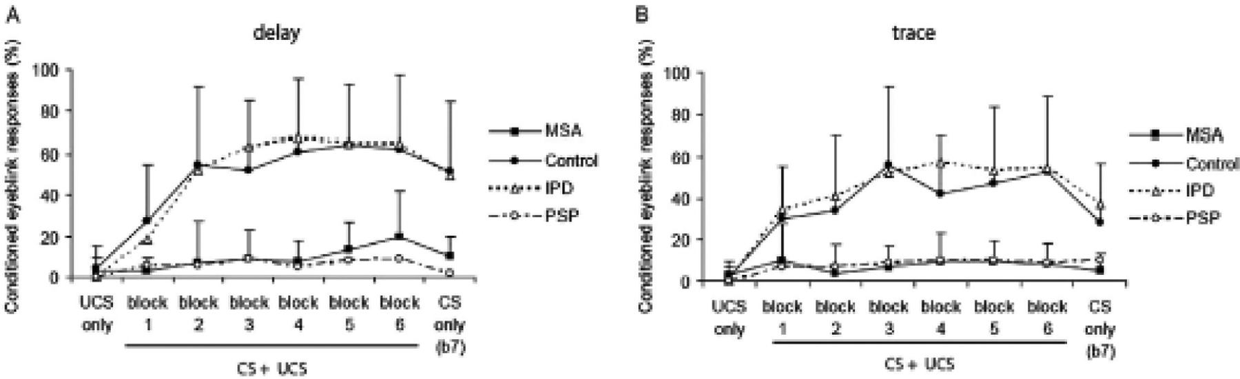
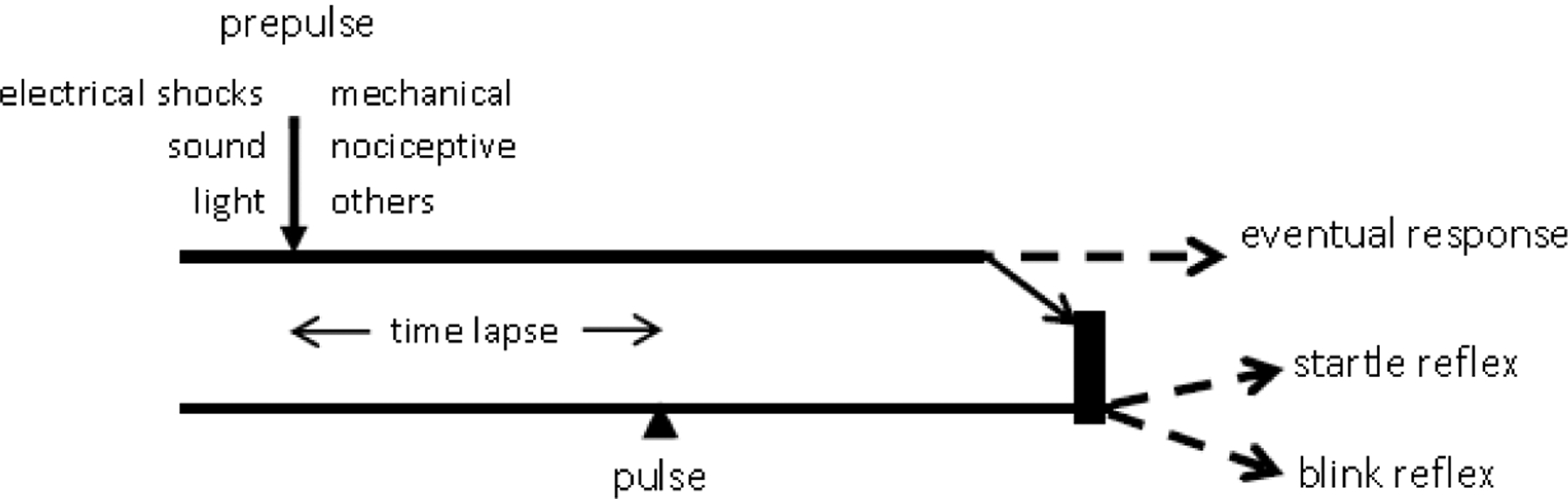
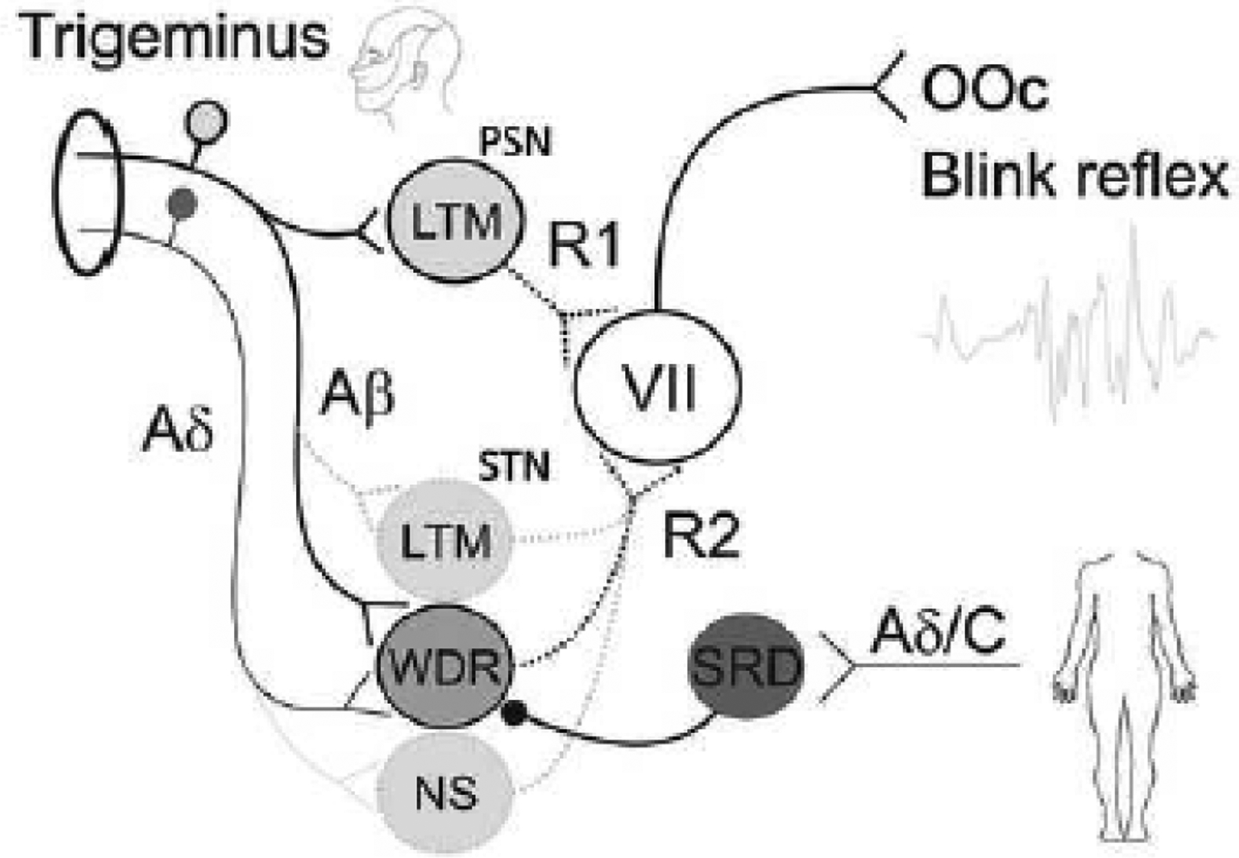
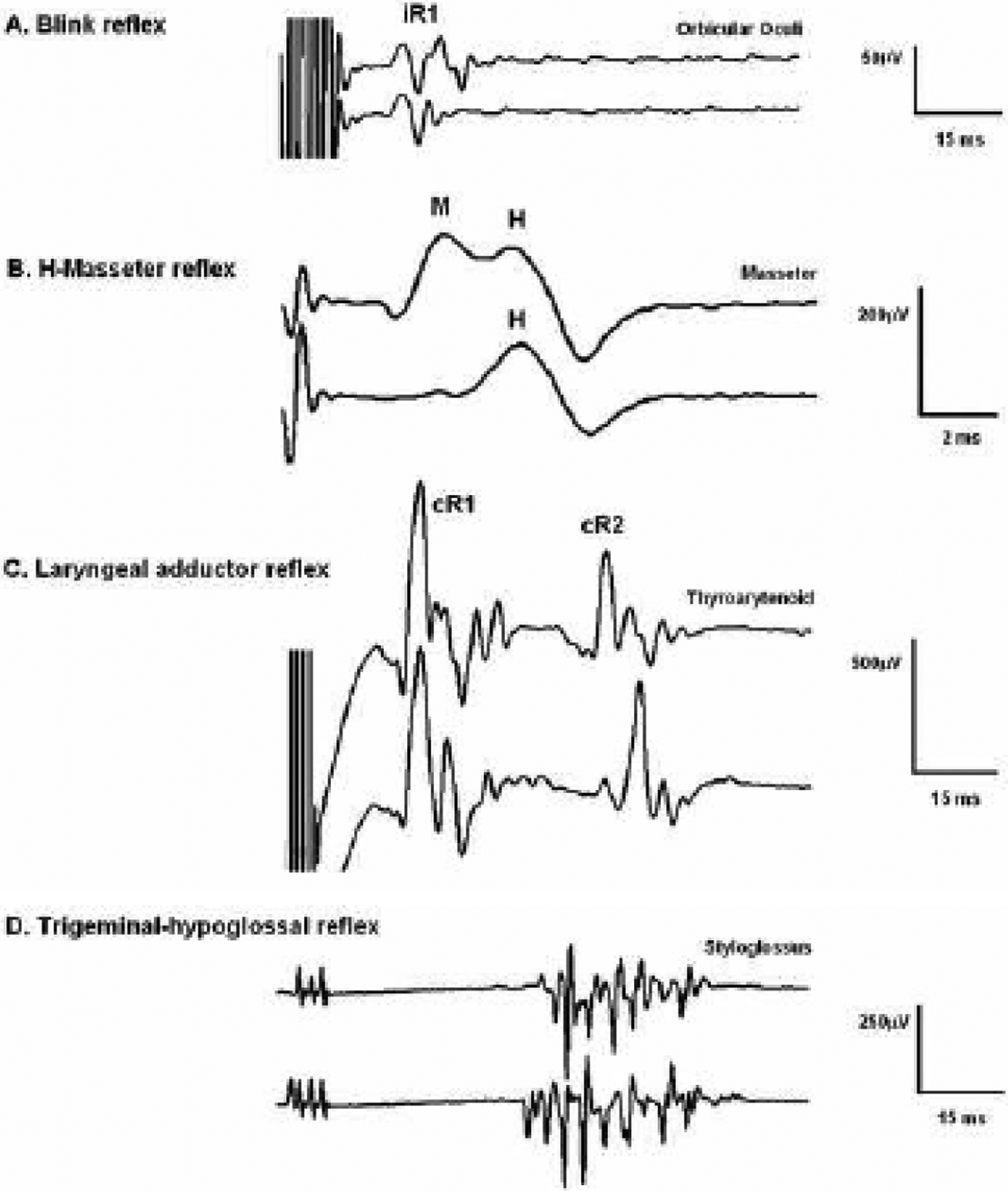
The blink reflex (BR) is a protective eye-closure reflex mediated by brainstem circuits. The BR is usually evoked by electrical supraorbital nerve stimulation but can be elicited by a variety of sensory modalities. It has a long history in clinical neurophysiology practice. Less is known, however, about the many ways to modulate the BR. Various neurophysiological techniques can be applied to examine different aspects of afferent and efferent BR modulation. In this line, classical conditioning, prepulse and paired-pulse stimulation, and BR elicitation by self-stimulation may serve to investigate various aspects of brainstem connectivity. The BR may be used as a tool to quantify top-down modulation based on implicit assessment of the value of blinking in a given situation, e.g., depending on changes in stimulus location and probability of occurrence. Understanding the role of non-nociceptive and nociceptive fibers in eliciting a BR is important to get insight into the underlying neural circuitry. Finally, the use of BRs and other brainstem reflexes under general anesthesia may help to advance our knowledge of the brainstem in areas not amenable in awake intact humans. This review summarizes talks held by the Brainstem Special Interest Group of the International Federation of Clinical Neurophysiology at the International Congress of Clinical Neurophysiology 2022 in Geneva, Switzerland, and provides a state-of-the-art overview of the physiology of BR modulation. Understanding the principles of BR modulation is fundamental for a valid and thoughtful clinical application (reviewed in part 2) (Gunduz et al., submitted).

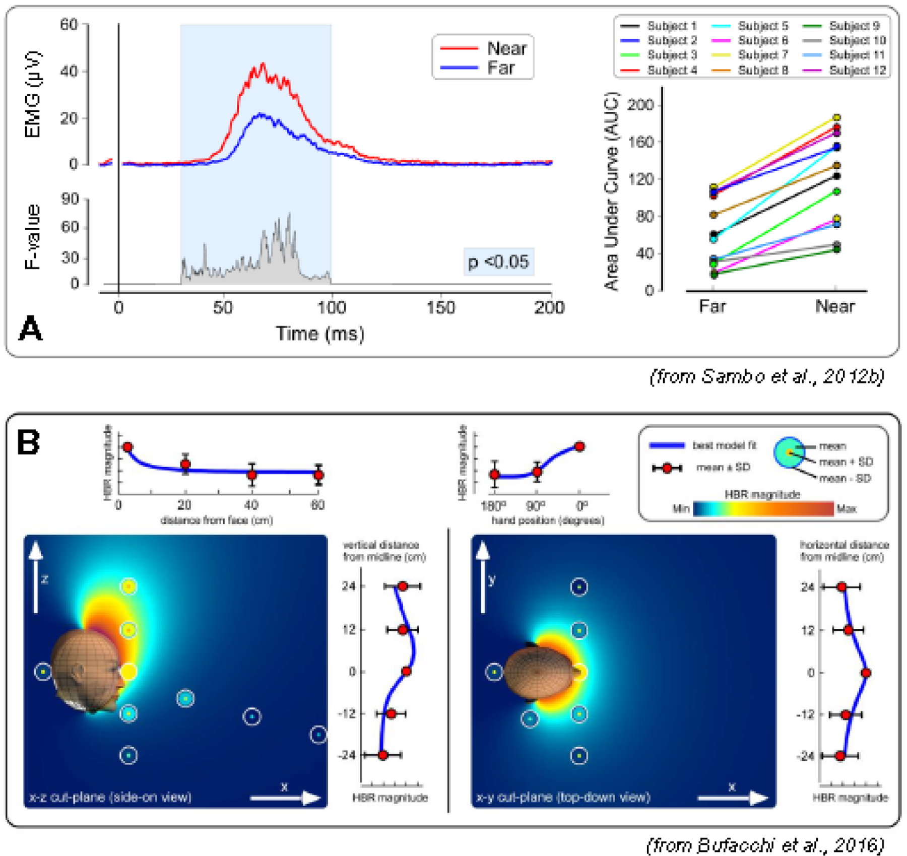
Keywords: Anesthesia; Classical conditioning; Pain; Peripersonal space; Prepulse inhibition; Protective reflex.


Conflict of interest statement

Conflict of interest MH: Relevant: none. Irrelevant: MH gets a share of patent royalties given to NIH by Brainsway for licensing a patent on the H-coil, a device for magnetic stimulation. He serves on the Medical Advisory Board of Brainsway, QuantalX and VoxNeuro. He has consulted for Janssen Pharmaceutical. None of the remaining authors has any conflict of interest to declare.

Figures

Figure 1.
Figure 1.
Simplified scheme of the blink reflex circuitry with the pontomedullary reticular formation as the central brainstem structure mediating all kinds of blink reflexes in response to various afferent modalities. CN = cochlear nuclei; PSN = principal sensory nucleus of the trigeminal nerve; STN = spinal trigeminal nucleus of the trigeminal nerve; VN = vestibular nuclei. Note that the early R1 response following supraorbital nerve (SON) stimulation is mediated via the PSN, connecting only with the ipsilateral facial nucleus, whereas the late responses are conveyed via the ipsilateral STN to the pontomedullary reticular formation from where ipsi- (R2) and contralateral (R2c) responses are generated. Blink reflexes evoked by other sensory modalities lack the ipsilateral R1 component. The reticular formation is a complex neuronal network containing numerous diffuse and highly organized regions (Crossman, 2005; Brodal, 2004c). Hence, only the main pathways are shown. Various afferents comprise the trigeminal nerve, in particular the SON (see section 2, Hopf, 1994; Pellegrini et al., 1995; Berardelli et al., 1999; Esteban, 1999; Aramideh and Ongerboer de Visser, 2002; Cruccu et al., 2005; Valls-Solé, 2012, 2019; Kimura, 2013), but also other branches of the trigeminal nerve (Kugelberg, 1952; Oka et al., 1958; Gandiglio and Fra, 1967; Kimura, 1973; Hess et al., 1984; Jääskeläinen, 1995; Valls-Solé et al., 1996; Pavesi et al., 1996). Extratrigeminal sensory afferents have been demonstrated by Gandiglio and Fra (1967), Miwa et al. (1995, 1996, 1998), and Alvarez-Blanco et al. (2009). Visual input is relayed via retinotectal and tectoreticular fibers (Crossman, 2005). Cochlear and vestibular afferents are described in Brodal’s chapters 8 and 9, respectively (Brodal, 2004a, 2004b) and in Gray’s Anatomy (Crossman, 2005). The pathway of the auditory blink reflex is depicted according to Hori et al. (1986), not showing the involvement of the colliculus inferior. The majority of trigeminal pain fibers, but not all, are transmitted through the STN (see section 7 for details).
Figure 2.
Figure 2.
Method of blink reflex conditioning. The horizontal axis is time with events marked. A. An unconditioned stimulus (US) produces an unconditioned response (UR). B. If a conditioned stimulus (CS) precedes the US multiple times, if there is learning, there will be a conditioned response (CR) coming near the time of the UR, usually just before it. B1 is trace conditioning with the CS ending before the delivery of the US (labelled CSt). B2 is delay conditioning with the CS ending at the same time as the US (labelled CSd). C. After successful conditioning, delivery of the CS will produce a CR even without any US. D. If the CS is delivered multiple times without the US, the CR will eventually be extinguished.
Figure 3.
Figure 3.
Delay conditioning in a control subject and a patient with cerebellar degeneration. Timing of the stimuli is noted in the lower left, CS is the conditioned stimulus and US is the unconditioned stimulus. The paradigm has 6 blocks with 10 trials per block, 8 with CS-US pairing, 1 with CS alone, and 1 with US alone, with a 10 second intertrial interval. The CS was a tone lasting 400 ms. Traces are rectified electromyographic activity in orbicularis oculi muscle. The first 6 traces are examples of responses to the pair of CS-US stimuli in the respective blocks, and the seventh trace is the CS, tone-alone, trace from the sixth block. In blocks 1 and 2 for the control subject and in all the blocks of the patient, the only response seen is the unconditioned response (UR). Conditioned responses (CR) can be seen preceding the UR in blocks 3 to 6 and the tone-alone block of the control subject. Only blinks in the interval between the vertical dotted lines are considered CR. Early blinks after delivery of the CS are called alpha blinks and are indicated with *. From Topka et al. (1993) with permission.
Figure 4.
Figure 4.
Blink reflex conditioning in idiopathic Parkinson’s disease (IPD), healthy controls, multiple system atrophy (MSA), and progressive supranuclear palsy (PSP). Delay conditioning on the left and trace conditioning on the right. The graphs show percent of conditioned responses (CR) in each of 7 blocks. The unconditioned stimulus (US) is a shock to the supraorbital nerve, the conditioned stimulus (CS) was a 400 ms tone. In delay conditioning the tone ended at the time of the shock; in trace conditioning there was a gap of 600 ms between the end of the tone and the shock. There were 6 blocks with pairing of US and CS in trials 1 to 9, an US only in trial 10, and a CS alone in trial 11. In the 7th block, there were 11 trials of the CS only (that would begin an extinction process). Note that conditioning is normal in IPD, but markedly diminished in MSA and PSP in both types of conditioning. From von Lewinski et al. (2013) with permission.
Figure 5.
Figure 5.
Simplified scheme of prepulse inhibition A prepulse stimulus of any of the modalities listed under ‘prepulse’ precedes a pulse stimulus, either a startling sound or a supraorbital nerve electrical stimulus, by a specific time lapse. The volley generated by the prepulse reaches the brainstem where it causes inhibition of the responses expected from the pulse stimulus, i.e., the startle reflex or the blink reflex (BR). The inhibition shows usually in a significant size reduction of either the startle reflex or the BR, depending on the pulse stimulus modality. The prepulse volley may also generate a response by itself, such as a BR, depending on the prepulse stimulus intensity. Similar mechanisms may apply in part also to circuits mentioned in Sections 5 and 6.
Figure 6.
Figure 6.
The magnitude of the blink reflex elicited by median nerve stimulation (hand-blink reflex, HBR) is modulated by the proximity between the stimulated hand and the face. A: the top waveforms are the rectified group average HBR for the hand position ”far” (blue) and “near” (red). The bottom waveform expresses the ANOVA F-value for each time point, in the significant time windows (P <0.05). The right panel shows a consistent effect across participants (single-subject HBR magnitudes are expressed as area under the curve, AUC) (modified from Sambo et al. (2012b)). B: geometric modelling of HBR strength as a function of stimulus position. Plots are a combined description of the experimental data with the bestfitting geometric model. Measured HBR data are represented as concentric circles located where the measurements were taken. Background color represents the HBR magnitude predicted by the best-fitting geometric model. Line graphs at the side of each color plot show HBR magnitudes along each axis, together with the best-fitting geometric model (blue line). HBR magnitude increases monotonically with the proximity between the stimulus and the face, and it is symmetrical on the axial plane, but asymmetrical along the rostro-caudal axis, with stronger HBR elicited by stimuli occurring above than below the face (from Bufacchi et al. (2016)).
Figure 7.
Figure 7.
Neuronal network model of the blink reflex (BR). The principal sensory nucleus (PSN) mediates the R1 component, and the spinal trigeminal nucleus (STN) mediates the R2 component of the BR. Trigeminal non-nociceptive and thick-myelinated Aβ afferents project on low-threshold mechanoreceptive (LTM) neurons of the PSN generating the R1 reflex of the orbicularis oculi muscle (OOc) via motor neurons of the facial nerve (VII). Trigeminal tactile Aβ and nociceptive Aδ afferents converge onto common wide-dynamic-range (WDR) interneurons of the STN generating the R2 reflex via motor neurons of the VII. Noxious stimulation to remote body sites such as the extremities, activate multireceptive neurons of the subnucleus reticularis dorsalis (SRD) inhibiting WDR neurons and, hence, the R2 reflex responses. Additionally, the R2 reflex may be evoked or modulated by Aβ fiber input on LTM neurons of the STN or noxious input from Aδ afferents on nociceptive specific neurons (NS) of the STN.
Figure 8.
Figure 8.
A. Blink reflex (BR) elicited in a patient under total intravenous anesthesia (TIVA, two trials shown). The BR was elicited by applying a short train of 9 electrical pulses (2 ms inter-stimulus-interval, at 36 mA intensity) to the supraorbital nerve, ipsilateral to the recorded side (Deletis et al., 2009). B. Laryngeal adductor reflex (LAR) elicited in a patient under TIVA (two trials shown). The LAR was elicited by applying a short train of 3 electrical pulses (1–2 ms inter-stimulus-interval, at 9 mA intensity) applied to the laryngeal mucosa contralateral to the recording side (Sinclair et al., 2017b). C. Masseter H reflex elicited in a patient under TIVA. The first trial was elicited by stimulating the third branch of the trigeminal nerve (under the zygomatic arc) with a single pulse at a slightly higher intensity than the second to show M/H and isolated H, respectively (Ulkatan et al., 2017). D. Trigeminal-hypoglossal reflex (THR) elicited in a patient under TIVA (two trials shown). The THR was elicited by stimulating the third branch of the trigeminal nerve (under the zygomatic arc) with a short train of 3 electrical pulses at 30 mA.

Similar articles

Cited by

References

    1. Accornero N, Berardelli A, Bini G, Cruccu G, Manfredi M. Corneal reflex elicited by electrical stimulation of the human cornea. Neurology 1980;30(7 Pt 1):782–5. 10.1212/wnl.30.7.782 - DOI - PubMed
    1. Accornero N, Berardelli A, Bini G, Cruccu G, Manfredi M, Caruso R, et al. [Method of recording the corneal reflex in humans, using physiological stimulation]. Boll Soc Ital Biol Sper 1978;54(3):249–51. - PubMed
    1. Adachi M, Nonaka S, Katada A, Arakawa T, Ota R, Harada H, et al. Carbachol injection into the pontine reticular formation depresses laryngeal muscle activities and airway reflexes in decerebrate cats. Neurosci Res 2010;67(1):40–50. 10.1016/j.neures.2010.01.009 - DOI - PubMed
    1. Alvarez-Blanco S, Leon L, Valls-Solé J. The startle reaction to somatosensory inputs: different response pattern to stimuli of upper and lower limbs. Exp Brain Res 2009;195(2):285–92. 10.1007/s00221-009-1784-7 - DOI - PubMed
    1. Ambalavanar R, Purcell L, Miranda M, Evans F, Ludlow CL. Selective suppression of late laryngeal adductor responses by N-methyl-D-aspartate receptor blockade in the cat. J Neurophysiol 2002;87(3):1252–62. 10.1152/jn.00595.2001 - DOI - PubMed

LinkOut - more resources