Profiling human brain vascular cells using single-cell transcriptomics and organoids
- PMID: 38102365
- PMCID: PMC11537086
- DOI: 10.1038/s41596-023-00929-1
Profiling human brain vascular cells using single-cell transcriptomics and organoids
Abstract
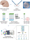
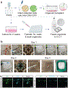
Angiogenesis and neurogenesis are functionally interconnected during brain development. However, the study of the vasculature has trailed other brain cell types because they are delicate and of low abundance. Here we describe a protocol extension to purify prenatal human brain endothelial and mural cells with FACS and utilize them in downstream applications, including transcriptomics, culture and organoid transplantation. This approach is simple, efficient and generates high yields from small amounts of tissue. When the experiment is completed within a 24 h postmortem interval, these healthy cells produce high-quality data in single-cell transcriptomics experiments. These vascular cells can be cultured, passaged and expanded for many in vitro assays, including Matrigel vascular tube formation, microfluidic chambers and metabolic measurements. Under these culture conditions, primary vascular cells maintain expression of cell-type markers for at least 3 weeks. Finally, we describe how to use primary vascular cells for transplantation into cortical organoids, which captures key features of neurovascular interactions in prenatal human brain development. In terms of timing, tissue processing and staining requires ~3 h, followed by an additional 3 h of FACS. The transplant procedure of primary, FACS-purified vascular cells into cortical organoids requires an additional 2 h. The time required for different transcriptomic and epigenomic protocols can vary based on the specific application, and we offer strategies to mitigate batch effects and optimize data quality. In sum, this vasculo-centric approach offers an integrated platform to interrogate neurovascular interactions and human brain vascular development.
© 2023. Springer Nature Limited.
Figures


References
-
- Paredes I, Himmels P & Ruiz de Almodóvar C Neurovascular Communication during CNS Development. Dev. Cell 45, 10–32 (2018). - PubMed
-
- Silva-Vargas V, Crouch EE & Doetsch F Adult neural stem cells and their niche: a dynamic duo during homeostasis, regeneration, and aging. Curr. Opin. Neurobiol 23, 935–942 (2013). - PubMed
-
- Gould DB et al. Mutations in Col4a1 cause perinatal cerebral hemorrhage and porencephaly. Science 308, 1167–1171 (2005). - PubMed
-
- Xu H. et al. Maturational changes in laminin, fibronectin, collagen IV, and perlecan in germinal matrix, cortex, and white matter and effect of betamethasone. J. Neurosci. Res 86, 1482–1500 (2008). - PubMed
Key references using this protocol:
-
- Crouch EE, Bhaduri A, Andrews MG, Cebrian-Silla A, Diafos LN, Birrueta JO, Wedderburn-Pugh K, Valenzuela EJ, Bennett NK, Eze UC, Sandoval-Espinosa C, Chen J, Mora C, Ross JM, Howard CE, Gonzalez-Granero S, Lozano JF, Vento M, Haeussler M, Paredes MF, Nakamura K, Garcia-Verdugo JM, Alvarez-Buylla A, Kriegstein AR, Huang EJ. Ensembles of endothelial and mural cells promote angiogenesis in prenatal human brain. Cell. 2022. Sep 29;185(20):3753–3769.e18. doi: 10.1016/j.cell.2022.09.004. - DOI - PMC - PubMed
Key data used in this protocol:
-
- Crouch EE, Bhaduri A, Andrews MG, Cebrian-Silla A, Diafos LN, Birrueta JO, Wedderburn-Pugh K, Valenzuela EJ, Bennett NK, Eze UC, Sandoval-Espinosa C, Chen J, Mora C, Ross JM, Howard CE, Gonzalez-Granero S, Lozano JF, Vento M, Haeussler M, Paredes MF, Nakamura K, Garcia-Verdugo JM, Alvarez-Buylla A, Kriegstein AR, Huang EJ. Ensembles of endothelial and mural cells promote angiogenesis in prenatal human brain. Cell. 2022. Sep 29;185(20):3753–3769.e18. doi: 10.1016/j.cell.2022.09.004. - DOI - PMC - PubMed
Publication types
MeSH terms
Associated data
- Actions
Grants and funding
- NS116161/U.S. Department of Health & Human Services | NIH | National Institute of Neurological Disorders and Stroke (NINDS)
- DK063720/U.S. Department of Health & Human Services | NIH | National Institute of Diabetes and Digestive and Kidney Diseases (National Institute of Diabetes & Digestive & Kidney Diseases)
- P30 DK063720/DK/NIDDK NIH HHS/United States
- NS083513/U.S. Department of Health & Human Services | NIH | National Institute of Neurological Disorders and Stroke (NINDS)
- U01 MH105989/MH/NIMH NIH HHS/United States
- K99 NS111731/NS/NINDS NIH HHS/United States
- K08 NS116161/NS/NINDS NIH HHS/United States
- K12 HD000850/HD/NICHD NIH HHS/United States
- SFSU EDUC2-12693/California Institute for Regenerative Medicine (CIRM)
- R00 NS111731/NS/NINDS NIH HHS/United States
- R00 MH125329/MH/NIMH NIH HHS/United States
- K99 MH125329/MH/NIMH NIH HHS/United States
- NS111731/U.S. Department of Health & Human Services | NIH | National Institute of Neurological Disorders and Stroke (NINDS)
- 857876/American Heart Association (American Heart Association, Inc.)
- no number/California Institute for Regenerative Medicine (CIRM)
- 5K12HD000850-34/U.S. Department of Health & Human Services | NIH | Eunice Kennedy Shriver National Institute of Child Health and Human Development (NICHD)
- MH125329/U.S. Department of Health & Human Services | NIH | National Institute of Mental Health (NIMH)
- MH105989/U.S. Department of Health & Human Services | NIH | National Institute of Mental Health (NIMH)
- P01 NS083513/NS/NINDS NIH HHS/United States

