. 2023 Dec 15;13(1):22302.
doi: 10.1038/s41598-023-49827-2.

Lipidomic assessment of the impact of Nannochloropsis oceanica microalga lipid extract on human skin keratinocytes exposed to chronic UVB radiation

Affiliations

Lipidomic assessment of the impact of Nannochloropsis oceanica microalga lipid extract on human skin keratinocytes exposed to chronic UVB radiation

Wojciech Łuczaj et al. Sci Rep. .

Abstract

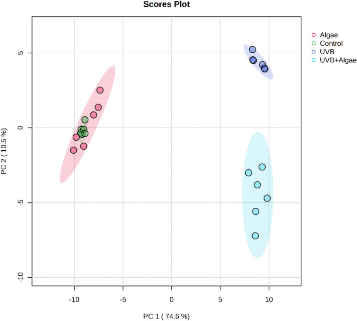
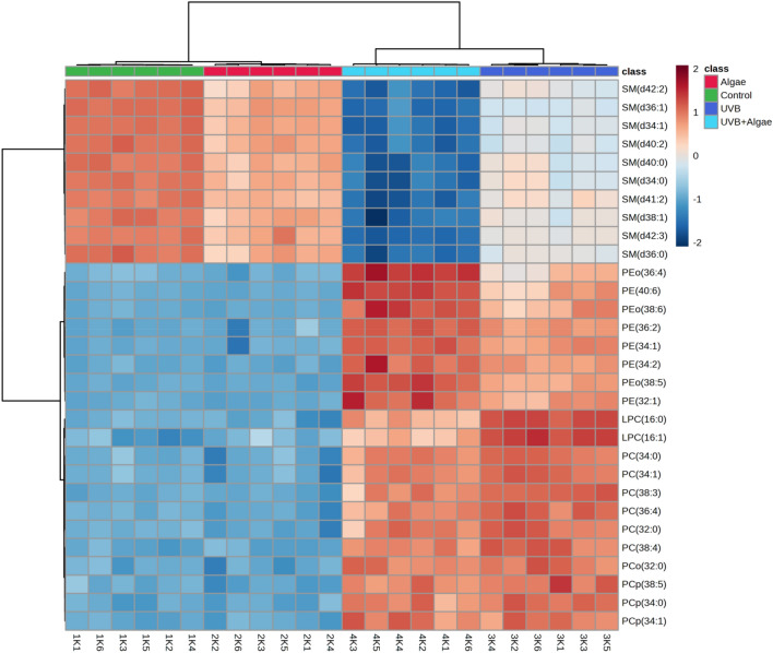
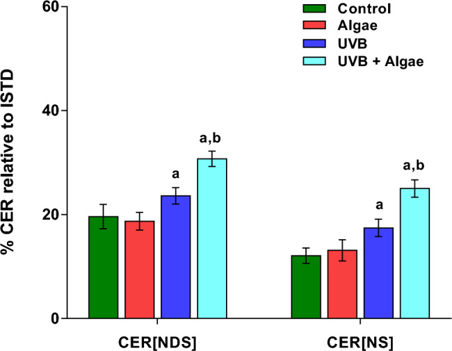
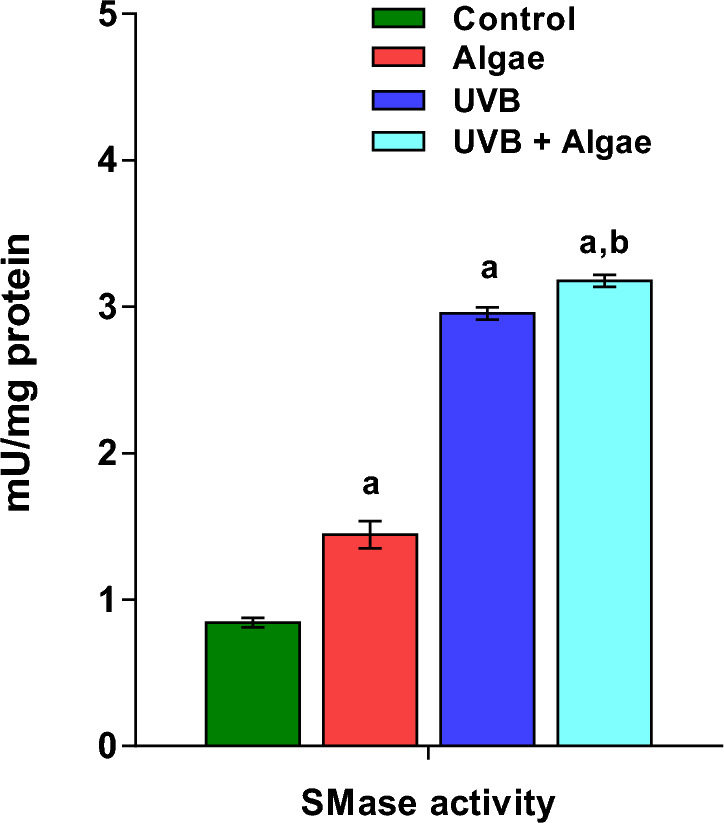
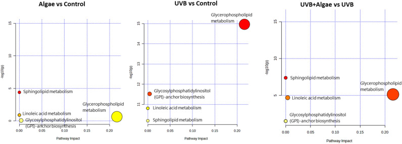
Considerable attention has been devoted to investigating the biological activity of microalgal extracts, highlighting their capacity to modulate cellular metabolism. This study aimed to assess the impact of Nannochloropsis oceanica lipid extract on the phospholipid profile of human keratinocytes subjected to UVB radiation. The outcomes revealed that treatment of keratinocytes with the lipid extract from microalgae led to a reduction in sphingomyelin (SM) levels, with a more pronounced effect observed in UVB-irradiated cells. Concomitantly, there was a significant upregulation of ceramides CER[NDS] and CER[NS], along with increased sphingomyelinase activity. Pathway analysis further confirmed that SM metabolism was the most significantly affected pathway in both non-irradiated and UVB-irradiated keratinocytes treated with the microalgal lipid extract. Additionally, the elevation in alkylacylPE (PEo) and diacylPE (PE) species content observed in UVB-irradiated keratinocytes following treatment with the microalgal extract suggested the potential induction of pro-survival mechanisms through autophagy in these cells. Conversely, a noteworthy reduction in LPC content in UVB-irradiated keratinocytes treated with the extract, indicated the anti-inflammatory properties of the lipid extract obtained from microalgae. However, to fully comprehend the observed alterations in the phospholipid profile of UVB-irradiated keratinocytes, further investigations are warranted to identify the specific fraction of compounds responsible for the activity of the Nannochloropsis oceanica extract.


Conflict of interest statement

The authors declare no competing interests.

Figures

Figure 1
Figure 1
Two-dimensional principal component analysis (2D PCA) scores plot of the relative phospholipid content in non-treated keratinocytes (Control), keratinocytes treated with an extract from the microalgae Nannochloropsis oceanica (3 µg/ml) [Algae], keratinocytes irradiated with UVB (60 mJ/cm2) [UVB] and keratinocytes irradiated with UVB (60 mJ/cm2) and treated with an extract from the microalgae Nannochloropsis oceanica (3 µg/ml) [UVB + Algae].
Figure 2
Figure 2
Two-dimensional hierarchical clustering heat map of the 30 main phospholipids of the four groups of keratinocytes, non-irradiated (Control), irradiated with UVB (UVB) and treated with an extract from the microalgae Nannochloropsis oceanica (Algae), and irradiated with UVB and treated with the extract (UVB + Algae), after the one-way ANOVA test. Levels of relative abundance are indicated on the color scale, with numbers indicating the fold difference from the grand mean. The clustering of the sample groups is represented by the dendrogram on the top. At the bottom appears the sample names. The clustering of individual phospholipid species with respect to their similarity in the change in relative abundance is shown by the dendrogram to the left.
Figure 3
Figure 3
Changes in relative ceramide content within the CER[NDS] and CER[NS] classes in the following keratinocyte groups: non-irradiated (Control), irradiated with UVB (UVB) and treated with an extract from the microalgae Nannochloropsis oceanica (Algae), and irradiated with UVB and treated with the extract (UVB + Algae). The presented values are expressed as mean ± SD; statistically significant differences (p < 0.05) in comparison to the: a—control group, b—UVB group.
Figure 4
Figure 4
Neutral sphingomyelinase (SMase) activity in the non-irradiated keratinocytes (Control), irradiated with UVB (UVB) and treated with an extract from the microalgae Nannochloropsis oceanica (Algae), and irradiated with UVB and treated with the extract (UVB + Algae). The presented values are expressed as mean ± SD; statistically significant differences (p < 0.05) in comparison to the: a—control group, b—UVB group.
Figure 5
Figure 5
Metabolic pathway analysis performed for the 30 most discriminating (according to One-way ANOVA) phospholipid species identified in keratinocytes treated with an extract from the microalgae Nannochloropsis oceanica (Algae) compared to non-treated keratinocytes (Control), and UVB-irradiated keratinocytes treated with extract from the microalgae Nannochloropsis oceanica (UVB + Algae) compared to cells irradiated with UVB (UVB). The colour of each circle is based on the p-value, while the size of the circle indicates pathway impact (a combination of the centrality and number of phospholipid species enriched in the pathway). Smaller p-values and larger pathway impact circles indicate a greater perturbation of the pathway. The y-axis represents the p-values from pathway enrichment analysis, while the x-axis represents pathway impact values from pathway topology analysis.

Similar articles

Cited by

References

    1. Ali A, Khan H, Bahadar R, Riaz A, Asad MHHB. The impact of airborne pollution and exposure to solar ultraviolet radiation on skin: Mechanistic and physiological insight. Environ. Sci. Pollut. Res. Int. 2020;27:28730–28736. doi: 10.1007/s11356-020-09280-4. - DOI - PubMed
    1. Panich U, Sittithumcharee G, Rathviboon N, Jirawatnotai S. Ultraviolet radiation-induced skin aging: The role of DNA damage and oxidative stress in epidermal stem cell damage mediated skin aging. Stem Cells Int. 2016;2016:7370642. doi: 10.1155/2016/7370642. - DOI - PMC - PubMed
    1. Stellavato, A. et al. Positive effects against UV-A induced damage and oxidative stress on an in vitro cell model using a hyaluronic acid based formulation containing amino acids, vitamins, and minerals. BioMed Res. Int. 2018, e8481243. https://www.hindawi.com/journals/bmri/2018/8481243/ (2018). - PMC - PubMed
    1. Zhang P, Wu MX. A clinical review of phototherapy for psoriasis. Lasers Med. Sci. 2018;33:173–180. doi: 10.1007/s10103-017-2360-1. - DOI - PMC - PubMed
    1. Sies H. Oxidative stress: Concept and some practical aspects. Antioxidants (Basel) 2020;9:852. doi: 10.3390/antiox9090852. - DOI - PMC - PubMed