

Radiomic features of the hippocampal based on magnetic resonance imaging in the menopausal mouse model linked to neuronal damage and cognitive deficits
- PMID: 38102441
- PMCID: PMC11156756
- DOI: 10.1007/s11682-023-00808-z
Radiomic features of the hippocampal based on magnetic resonance imaging in the menopausal mouse model linked to neuronal damage and cognitive deficits
Abstract
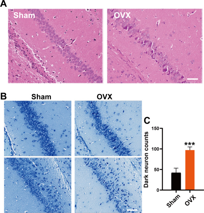
Estrogen deficiency in the early postmenopausal phase is associated with an increased long-term risk of cognitive decline or dementia. Non-invasive characterization of the pathological features of the pathological hallmarks in the brain associated with postmenopausal women (PMW) could enhance patient management and the development of therapeutic strategies. Radiomics is a means to quantify the radiographic phenotype of a diseased tissue via the high-throughput extraction and mining of quantitative features from images acquired from modalities such as CT and magnetic resonance imaging (MRI). This study set out to explore the correlation between radiomics features based on MRI and pathological features of the hippocampus and cognitive function in the PMW mouse model. Ovariectomized (OVX) mice were used as PWM models. MRI scans were performed two months after surgery. The brain's hippocampal region was manually annotated, and the radiomic features were extracted with PyRadiomics. Chemiluminescence was used to evaluate the peripheral blood estrogen level of mice, and the Morris water maze test was used to evaluate the cognitive ability of mice. Nissl staining and immunofluorescence were used to quantify neuronal damage and COX1 expression in brain sections of mice. The OVX mice exhibited marked cognitive decline, brain neuronal damage, and increased expression of mitochondrial complex IV subunit COX1, which are pathological phenomena commonly observed in the brains of AD patients, and these phenotypes were significantly correlated with radiomics features (p < 0.05, |r|>0.5), including Original_firstorder_Interquartile Range, Original_glcm_Difference Average, Original_glcm_Difference Average and Wavelet-LHH_glszm_Small Area Emphasis. Meanwhile, the above radiomics features were significantly different between the sham-operated and OVX groups (p < 0.01) and were associated with decreased serum estrogen levels (p < 0.05, |r|>0.5). This initial study indicates that the above radiomics features may have a role in the assessment of the pathology of brain damage caused by estrogen deficiency using routinely acquired structural MR images.
Keywords: Cognitive decline; Estrogen deficiency; MRI; Ovariectomized mouse; Radiomics features.
© 2023. The Author(s).
Conflict of interest statement
The authors declare no conflict of interest.
Figures


References
-
- Bittar, A., Al-Lahham, R., Bhatt, N., Moore, K., Montalbano, M., Jerez, C., Fung, L., McAllen, S., Ellsworth, A., & Kayed, R. (2022). Passive Immunotherapy Targeting Tau Oligomeric Strains Reverses Tauopathy Phenotypes in Aged Human-Tau Mice in a Mouse Model-Specific Manner. Journal of Alzheimer’s disease: JAD. - PubMed
-
- Cai J, Zheng J, Shen J, Yuan Z, Xie M, Gao M, Tan H, Liang Z, Rong X, Li Y, et al. A Radiomics Model for Predicting the response to Bevacizumab in Brain Necrosis after Radiotherapy. Clinical cancer Research: An Official Journal of the American Association for Cancer Research. 2020;26:5438–5447. doi: 10.1158/1078-0432.CCR-20-1264. - DOI - PubMed
MeSH terms
Substances
Grants and funding
LinkOut - more resources
Full Text Sources
Medical
