. 2023 Dec 15;11(1):93.
doi: 10.1186/s40635-023-00580-w.

Pressure-support compared with pressure-controlled ventilation mitigates lung and brain injury in experimental acute ischemic stroke in rats

Affiliations

Pressure-support compared with pressure-controlled ventilation mitigates lung and brain injury in experimental acute ischemic stroke in rats

Adriana L da Silva et al. Intensive Care Med Exp. .

Abstract

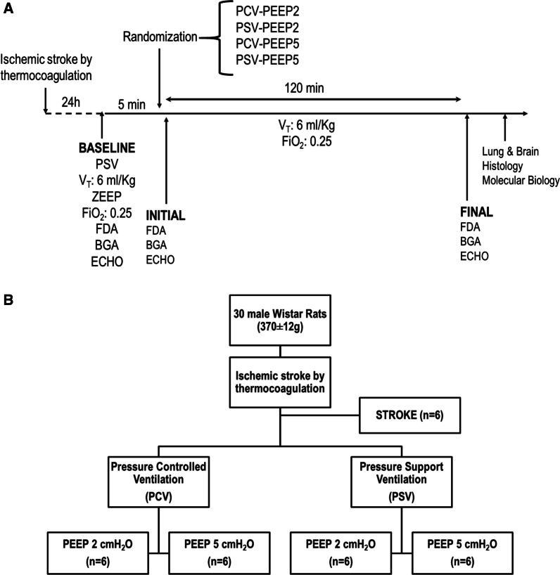
Background: We aimed to evaluate the pulmonary and cerebral effects of low-tidal volume ventilation in pressure-support (PSV) and pressure-controlled (PCV) modes at two PEEP levels in acute ischemic stroke (AIS).

Methods: In this randomized experimental study, AIS was induced by thermocoagulation in 30 healthy male Wistar rats. After 24 h, AIS animals were randomly assigned to PSV or PCV with VT = 6 mL/kg and PEEP = 2 cmH2O (PSV-PEEP2 and PCV-PEEP2) or PEEP = 5 cmH2O (PSV-PEEP5 and PCV-PEEP5) for 2 h. Lung mechanics, arterial blood gases, and echocardiography were evaluated before and after the experiment. Lungs and brain tissue were removed for histologic and molecular biology analysis. The primary endpoint was diffuse alveolar damage (DAD) score; secondary endpoints included brain histology and brain and lung molecular biology markers.

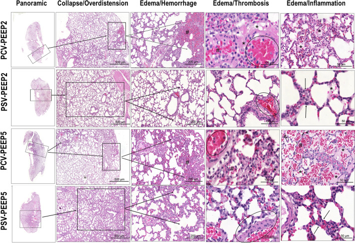
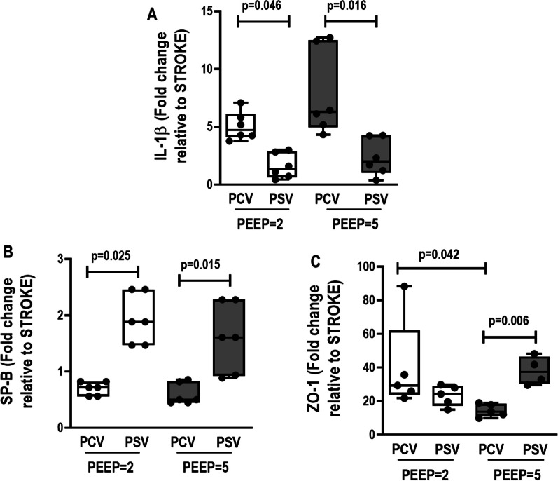
Results: In lungs, DAD was lower with PSV-PEEP5 than PCV-PEEP5 (p < 0.001); interleukin (IL)-1β was lower with PSV-PEEP2 than PCV-PEEP2 (p = 0.016) and PSV-PEEP5 than PCV-PEEP5 (p = 0.046); zonula occludens-1 (ZO-1) was lower in PCV-PEEP5 than PCV-PEEP2 (p = 0.042). In brain, necrosis, hemorrhage, neuropil edema, and CD45 + microglia were lower in PSV than PCV animals at PEEP = 2 cmH2O (p = 0.036, p = 0.025, p = 0.018, p = 0.011, respectively) and PEEP = 5 cmH2O (p = 0.003, p = 0.003, p = 0.007, p = 0.003, respectively); IL-1β was lower while ZO-1 was higher in PSV-PEEP2 than PCV-PEEP2 (p = 0.009, p = 0.007, respectively), suggesting blood-brain barrier integrity. Claudin-5 was higher in PSV-PEEP2 than PSV-PEEP5 (p = 0.036).

Conclusion: In experimental AIS, PSV compared with PCV reduced lung and brain injury. Lung ZO-1 reduced in PCV with PEEP = 2 versus PEEP = 5 cmH2O, while brain claudin-5 increased in PSV with PEEP = 2 versus PEEP = 5 cmH2O.

Keywords: Acute ischemic stroke; Brain injury; Lung injury; Mechanical ventilation.


Conflict of interest statement

The authors declare that they have no competing interests.

Figures

Fig. 1
Fig. 1
A Timeline representation of the experimental protocol. Respiratory system mechanics, arterial blood gases, and echocardiography were evaluated at the INITIAL and FINAL time points. B Schematic flowchart of the study design. BGA, blood gas analysis; ECHO, echocardiography; FDA, functional data acquisition; FiO2, fraction of inspired oxygen; PCV, pressure-controlled ventilation; PEEP, positive end-expiratory pressure; PSV, pressure-support ventilation; VT, tidal volume
Fig. 2
Fig. 2
Histoarchitecture of the lung. Representative images stained with hematoxylin and eosin (HE) according to diffuse alveolar damage (DAD) features: collapse/overdistension; edema/hemorrhage; edema/thrombosis; edema/inflammation. PCV, pressure-controlled ventilation; PEEP, positive end-expiratory pressure; PSV, pressure-support ventilation
Fig. 3
Fig. 3
Expression of biological markers in the lung related to inflammation (interleukin [IL]-1β), epithelial cell damage (surfactant protein-B [SP-B]), and endothelial cell damage (zonula occludens-1 [ZO-1]). Boxes show the interquartile range (25% to 75%), whiskers encompass the range (minimum to maximum), and horizontal lines represent median values of 6 animals/group. Comparisons were done by Kruskal–Wallis test followed by Dunn’s multiple comparisons test (p < 0.05)
Fig. 4
Fig. 4
Histoarchitecture of the hypothalamus. Visualized at low magnification stained by hematoxylin and eosin and at high magnification immunostained for CD11b and CD45 in PCV-PEEP2, PSV-PEEP2, PCV-PEEP5, and PSV-PEEP5. PCV-PEEP2 and PCV-PEEP5 exhibiting necrosis (*) and hemorrhage (#) in the hippocampus distorting the curved histoarchitecture (a, lateral) of the pyramidal cells band encircling the dentate gyrus (b, lateral) and obstructing the choroid plexus. At high magnification, numerous CD11b + mononuclear phagocytes visualized in microglia (thin arrows) adjacent to the dentate gyrus (DG) of hippocampus parenchyma, and numerous CD45 + cells in the choroid plexus (CP, thick arrows), representing mainly myeloid-derived macrophages that reside at the interfaces of the brain and periphery. Compared with the PCV-PEEP5 group, the number of microglia and CD11b + myeloid cells, as well as choroid plexus CD45 +, was more abundant in the PCV-PEEP2 group in the three brain compartments of the hippocampus parenchyma including the DG, pyramidal neurons, and CP (arrows). Note the intense edema of the neuropils (NP) in the PCV group. PSV-PEEP2 and PSV-PEEP5 showing preserved hippocampus histoarchitecture as a multiply curved structure (a, middle, bottom) with pyramidal cells forming a band that circles the denser line of small cells comprising the DG (b, top) and CP (c, center). At high magnification, CD11b + cells comprised mononuclear phagocytes, which were divided further into two populations: CD11b + cells characterized by microglia (thin arrows) that reside in the DG of hippocampus parenchyma, and CD45 + cells that reside in the CP (thick arrows). They represented mainly myeloid-derived macrophages that reside at the interfaces of the brain and periphery. Compared with the PSV-PEEP5 group, the numbers of microglia and CD11b + myeloid cells, as well as Cp CD45 +, were more prominent in the PCV-P2 group in the three brain compartments of the hippocampus parenchyma, including the DG, pyramidal neurons and CP. PCV, pressure-controlled ventilation; PEEP, positive end-expiratory pressure; PSV, pressure-support ventilation
Fig. 5
Fig. 5
Expression of biological markers in the brain related to neuroinflammation (interleukin [IL]-1β), and endothelial cell damage (claudin-5 and zonula occludens-1 [ZO-1]) in the following groups: NV, nonventilated; PCV, pressure-controlled ventilation; PSV, pressure-support ventilation; PEEP, positive end-expiratory pressure. Relative gene expression was calculated as a ratio of the average gene expression levels compared with the reference gene (acidic ribosomal phosphoprotein P0 [36B4]) and expressed as fold change relative to nonventilated animals (NV). Boxes show the interquartile range (25–75%), whiskers encompass the range (minimum to maximum), and horizontal lines represent median values of 6 animals/group. Comparisons were done by Kruskal–Wallis test followed by Dunn’s multiple comparisons test (p < 0.05)
Fig. 6
Fig. 6
Cardiac output and brain damage correlation. Cardiac output was negatively correlated with interleukin (IL)-1β (r = − 0.46, p = 0.023) positively associated with Zonula occludens (ZO)-1 gene expression at brain tissue (r = 0.38, p = 0.068)

Similar articles

Cited by

References

    1. Robba C, Poole D, McNett M, Asehnoune K, Bosel J, Bruder N, et al. Mechanical ventilation in patients with acute brain injury: recommendations of the European Society of Intensive Care Medicine consensus. Intensive Care Med. 2020;46(12):2397–2410. doi: 10.1007/s00134-020-06283-0. - DOI - PMC - PubMed
    1. Bruni A, Garofalo E, Pelaia C, Longhini F, Navalesi P. Mechanical ventilation in brain injured patients: seeing the forest for the trees. J Thorac Dis. 2017;9(10):3483–3487. doi: 10.21037/jtd.2017.08.149. - DOI - PMC - PubMed
    1. Asehnoune K, Mrozek S, Perrigault PF, Seguin P, Dahyot-Fizelier C, Lasocki S, et al. A multi-faceted strategy to reduce ventilation-associated mortality in brain-injured patients. The BI-VILI project: a nationwide quality improvement project. Intensive Care Med. 2017;43(7):957–970. doi: 10.1007/s00134-017-4764-6. - DOI - PubMed
    1. Fan TH, Huang M, Gedansky A, Price C, Robba C, Hernandez AV, et al. Prevalence and outcome of acute respiratory distress syndrome in traumatic brain injury: a systematic review and meta-analysis. Lung. 2021;199(6):603–610. doi: 10.1007/s00408-021-00491-1. - DOI - PMC - PubMed
    1. Greenberg SM, Ziai WC, Cordonnier C, Dowlatshahi D, Francis B, Goldstein JN, et al. 2022 Guideline for the Management of Patients With Spontaneous Intracerebral Hemorrhage: a Guideline From the American Heart Association/American Stroke Association. Stroke. 2022;53(7):e282–e361. doi: 10.1161/STR.0000000000000407. - DOI - PubMed