An MRI-based radiomics nomogram for detecting cervical esophagus invasion in hypopharyngeal squamous cell carcinoma
- PMID: 38102719
- PMCID: PMC10724942
- DOI: 10.1186/s40644-023-00642-y
An MRI-based radiomics nomogram for detecting cervical esophagus invasion in hypopharyngeal squamous cell carcinoma
Abstract
Background: Accurate detection of cervical esophagus invasion (CEI) in HPSCC is challenging but crucial. We aimed to investigate the value of magnetic resonance imaging (MRI)-based radiomics for detecting CEI in patients with HPSCC.
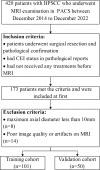
Methods: This retrospective study included 151 HPSCC patients with or without CEI, which were randomly assigned into a training (n = 101) or validation (n = 50) cohort. A total of 750 radiomics features were extracted from T2-weighted imaging (T2WI) and contrast-enhanced T1-weighted imaging (ceT1WI), respectively. A radiomics signature was constructed using the least absolute shrinkage and selection operator method. Multivariable logistic regression analyses were adopted to establish a clinical model and a radiomics nomogram. Two experienced radiologists evaluated the CEI status based on morphological findings. Areas under the curve (AUCs) of the models and readers were compared using the DeLong method. The performance of the nomogram was also assessed by its calibration and clinical usefulness.
Results: The radiomics signature, consisting of five T2WI and six ceT1WI radiomics features, was significantly associated with CEI in both cohorts (all p < 0.001). The radiomics nomogram combining the radiomics signature and clinical T stage achieved significantly higher predictive value than the clinical model and pooled readers in the training (AUC 0.923 vs. 0.723 and 0.621, all p < 0.001) and validation (AUC 0.888 vs. 0.754 and 0.647, all p < 0.05) cohorts. The radiomics nomogram showed favorable calibration in both cohorts and provided better net benefit than the clinical model.
Conclusions: The MRI-based radiomics nomogram is a promising method for detecting CEI in HPSCC.
Keywords: Cervical esophagus invasion; Hypopharyngeal squamous cell carcinoma; Magnetic resonance imaging; Radiomics.
© 2023. The Author(s).
Conflict of interest statement
The authors declare no conflict of interest.
Figures


Similar articles
-
An MRI-based radiomics-clinical nomogram for the overall survival prediction in patients with hypopharyngeal squamous cell carcinoma: a multi-cohort study.Eur Radiol. 2022 Mar;32(3):1548-1557. doi: 10.1007/s00330-021-08292-z. Epub 2021 Oct 19. Eur Radiol. 2022. PMID: 34665315
-
Computed tomography-based absolute delta radiomics nomogram for predicting perineural invasion in hypopharyngeal squamous cell carcinoma.Eur J Radiol. 2025 Feb;183:111912. doi: 10.1016/j.ejrad.2024.111912. Epub 2025 Jan 5. Eur J Radiol. 2025. PMID: 39809043
-
MRI-based radiomics nomogram for distinguishing solitary fibrous tumor from schwannoma in the orbit: a two-center study.Eur Radiol. 2024 Jan;34(1):560-568. doi: 10.1007/s00330-023-10031-5. Epub 2023 Aug 3. Eur Radiol. 2024. PMID: 37532903 Clinical Trial.
-
Multiparametric MRI-based radiomics nomogram for preoperative prediction of lymphovascular invasion and clinical outcomes in patients with breast invasive ductal carcinoma.Eur Radiol. 2022 Jun;32(6):4079-4089. doi: 10.1007/s00330-021-08504-6. Epub 2022 Jan 20. Eur Radiol. 2022. PMID: 35050415
-
Radiomics in Hypopharyngeal Cancer Management: A State-of-the-Art Review.Biomedicines. 2023 Mar 6;11(3):805. doi: 10.3390/biomedicines11030805. Biomedicines. 2023. PMID: 36979783 Free PMC article. Review.
References
-
- Amin MB, Greene FL, Edge SB, Compton CC, Gershenwald JE, Brookland RK, Meyer L, Gress DM, Byrd DR, Winchester DP. The Eighth Edition AJCC Cancer staging Manual: continuing to build a bridge from a population-based to a more personalized approach to cancer staging. Cancer J Clin. 2017;67:93–9. doi: 10.3322/caac.21388. - DOI - PubMed
MeSH terms
Grants and funding
LinkOut - more resources
Full Text Sources
Medical

