. 2023 Dec 15;42(1):342.
doi: 10.1186/s13046-023-02928-2.

RGCC-mediated PLK1 activity drives breast cancer lung metastasis by phosphorylating AMPKα2 to activate oxidative phosphorylation and fatty acid oxidation

Affiliations

RGCC-mediated PLK1 activity drives breast cancer lung metastasis by phosphorylating AMPKα2 to activate oxidative phosphorylation and fatty acid oxidation

Shaojie Cheng et al. J Exp Clin Cancer Res. .

Abstract

Background: More than 90% of the mortality of triple-negative breast cancer (TNBC) patients is attributed to cancer metastasis with organotropism. The lung is a frequent site of TNBC metastasis. However, the precise molecular mechanism for lung-specific metastasis of TNBC is not well understood.

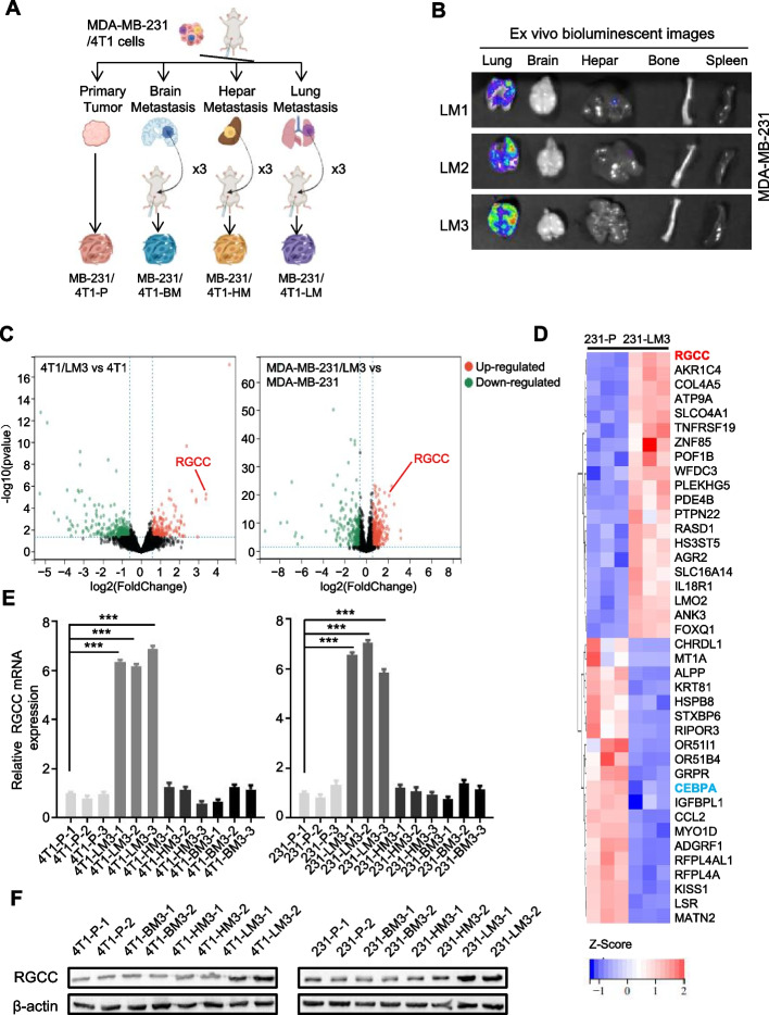
Methods: RNA sequencing was performed to identify patterns of gene expression associated with lung metastatic behavior using 4T1-LM3, MBA-MB-231-LM3, and their parental cells (4T1-P, MBA-MB-231-P). Expressions of RGCC, called regulator of cell cycle or response gene to complement 32 protein, were detected in TNBC cells and tissues by qRT-PCR, western blotting, and immunohistochemistry. Kinase activity assay was performed to evaluate PLK1 kinase activity. The amount of phosphorylated AMP-activated protein kinase α2 (AMPKα2) was detected by immunoblotting. RGCC-mediated metabolism was determined by UHPLC system. Oxidative phosphorylation was evaluated by JC-1 staining and oxygen consumption rate (OCR) assay. Fatty acid oxidation assay was conducted to measure the status of RGCC-mediated fatty acid oxidation. NADPH and ROS levels were detected by well-established assays. The chemical sensitivity of cells was evaluated by CCK8 assay.

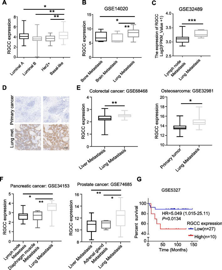
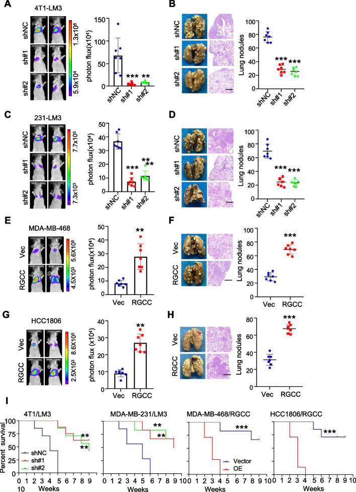
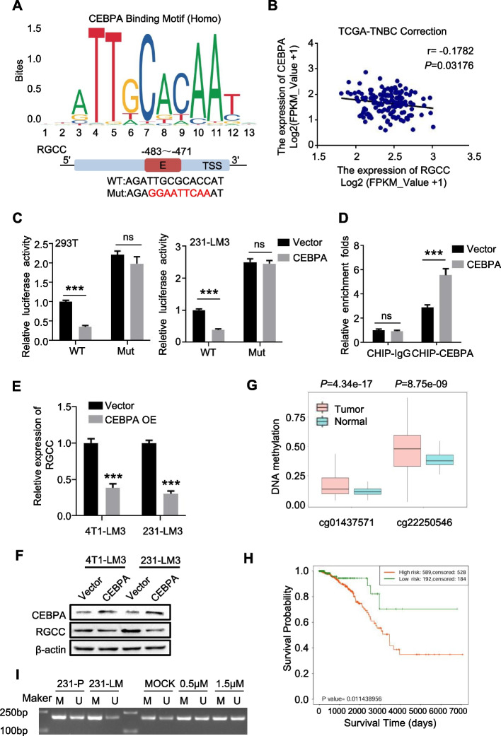
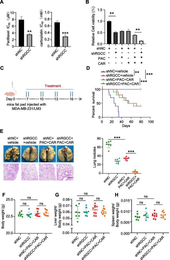
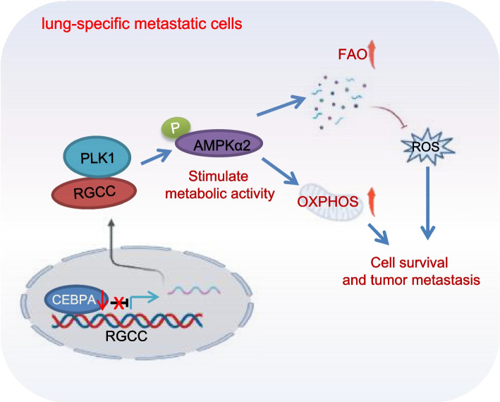
Results: RGCC is aberrantly upregulated in pulmonary metastatic cells. High level of RGCC is significantly related with lung metastasis in comparison with other organ metastases. RGCC can effectively promote kinase activity of PLK1, and the activated PLK1 phosphorylates AMPKα2 to facilitate TNBC lung metastasis. Mechanistically, the RGCC/PLK1/AMPKα2 signal axis increases oxidative phosphorylation of mitochondria to generate more energy, and promotes fatty acid oxidation to produce abundant NADPH. These metabolic changes contribute to sustaining redox homeostasis and preventing excessive accumulation of potentially detrimental ROS in metastatic tumor cells, thereby supporting TNBC cell survival and colonization during metastases. Importantly, targeting RGCC in combination with paclitaxel/carboplatin effectively suppresses pulmonary TNBC lung metastasis in a mouse model.

Conclusions: RGCC overexpression is significantly associated with lung-specific metastasis of TNBC. RGCC activates AMPKα2 and downstream signaling through RGCC-driven PLK1 activity to facilitate TNBC lung metastasis. The study provides implications for RGCC-driven OXPHOS and fatty acid oxidation as important therapeutic targets for TNBC treatment.

Keywords: Fatty acid oxidation; Lung metastasis; OXPHOS; PLK1; RGCC.


Conflict of interest statement

The authors declare that they have no competing interests.

Figures

Fig. 1
Fig. 1
The enhanced RGCC in lung metastatic TNBC cells. A Schematic diagram to show the organotropic metastasis mouse model establishment of TNBC cells. B Representative bioluminescence images of lung-tropic metastasis of MDA-MB-231 cells in major organs. C Volcano plots of the altered gene expression between lung metastatic cells (LM3) and parental cells (P). The red and green dots represent the significantly upregulated and downregulated RNAs, respectively. D Heatmap shows 20 most upregulated or downregulated genes between MDA-MB-231/LM3 and MDA-MB-231/P cells. E Relative folds of RGCC mRNA expression in various metastatic organ sites were detected by qRT-PCR (P: parental; LM3/HM3/BM3: the third lung/liver/brain metastatic cells.). F Western blot analysis of RGCC protein levels in metastatic 4T1/MDA-MB-231 derived from various organ lesion and parental cells. (The data are presented as the mean ± SD; ***P < 0.001)
Fig. 2
Fig. 2
The increased RGCC is related with TNBC clinical features. A-C Analysis of RGCC expression in human breast cancer subtypes (A) and BC patients with different organ metastases (B, C) based on datasets from the TCGA database. In the boxplot, the middle line represents the median, and the bottom and top line correspond to the 25th and 75th percentiles. D Representative IHC images of RGCC in clinical tumor tissues of TNBC in site and lung metastases (Scale bars, 200 μm). E, F Increased RGCC mRNA expression was in lung metastases compared with other organs metastases or primary tumor based on the analysis of microarray datasets from different metastatic solid tumors. G Kaplan–Meier analysis to show patients’ survival of TNBC with high or low level of RGCC. (*P < 0.05, **P < 0.01, ***P < 0.001)
Fig. 3
Fig. 3
RGCC is a promoter for TNBC lung metastasis. A-D Intravenous injection of indicated TNBC cells with RGCC knockdown or control cells for 30 days, representative bioluminescence imaging (BLI) of lung metastasis (A, C) (n = 7 mice per group), pulmonary surface nodules, H&E images (B, D) (n = 7 mice per group) were shown (scale bars, 200 μm). E–H Experiments described as above, BLI (E) (n = 7 mice per group) (E, G), pulmonary surface nodules, H&E images (F, H) (n = 7 mice per group) in mice injected indicated TNBC cells with or without ectopic RGCC (scale bars, 400 μm). I Kaplan–Meier survival analyses of TNBC metastatic mice indicated as A-H (n = 7 mice per group). (The data are presented as the mean ± SD; **P < 0.01, ***P < 0.001)
Fig. 4
Fig. 4
CEBPA negatively regulates RGCC expression. A The recognition motif of CEBPA in RGCC promoter (upper) analyzed using the JASPAR database, and the schematic illustration (down) showing the potential CEBPA responsive elements (E1). B A negative correlation between RGCC and CEBPA expression by Pearson correlation analysis using TCGA-TNBC dataset. C Luciferase reporter assays showing regulation of CEBPA regulation of RGCC promoter activity in 293 T and MDA-MB-231/LM3 cells. D ChIP assays to verify CEBPA binding affinity to the RGCC promoter in MDA-MB-231/LM3 transfected with CEBPA or control vector. E, F The regulation of CEBPA to RGCC was determined by qRT-PCR (E) and western blotting (F) in lung metastatic 4T1/LM3 and MDA-MB-231/LM3 cells. G High level of methylated CEBPA was in breast tumors than in normal tissues. H The Kaplan–Meier survival curves of BC patients with high or low methylated CEBPA. I Detection of methylation level in CpG islands of the CEBPA promoter by methylation specific polymerase chain reaction (MSP) (M: Methylation primer, U: Unmethylation primer.). (The data are presented as the mean ± SD; *P < 0.05, **P < 0.01, ***P < 0.001)
Fig. 5
Fig. 5
RGCC stimulates PLK1 kinase activity. A Venn diagram showing the potential interacting proteins of RGCC predicted by the database of Inbio discover, String and Biogrid. B Identification of the potential proteins interacting with RGCC by Mass Spectrometry. C Co-IP assays to confirm the direct interaction between RGCC and PLK1 in MDA-MB-231/LM3 cells using antibodies anti-RGCC or anti-PLK1, respectively. D IF co-staining showing the co-localization of RGCC (Red) and PLK1 (Green) in 4T1/LM3 and MDA-MB-231/LM3 cells. E Co-immunoprecipitation assay was used to detect the direct binding domain using HA-RGCC and FLAG-PLK1 truncation fragments in 293 T cells. F The binding mode of RGCC and PLK1 kinase domain (KD). G Western blot analysis of PLK1 protein levels under RGCC knockdown or overexpression. H PLK1 kinase assay showing RGCC affects PLK1 kinase activity. Data are means ± SD from 3 independent experiments
Fig. 6
Fig. 6
The RGCC/PLK1 axis activates AMPKα2. A The significant pathways associated with PLK1 were enriched by SangerBox based on the PLK1 potential phosphorylated proteins. B Determination of AMPKα2 phosphorylation in vitro. The experiments were independently repeated three times, with the error bars denoting the S.D. C Western blots of p-AMPK and p-ACC proteins in RGCC knockdown or overexpression tumor cells. D Orthotopic metastasis of RGCC overexpressed MDA-MB-468 cells treated with or without PLK1 inhibitor (Volasertb) or AMPK inhibitor (Dorsomorphin). Volasertib at 25 mg/kg, dorsomorphin at 0.2 mg/kg, or O304 at 200 mg/kg were given twice per week by oral gavage after the mouse’s tumor volume was above 50 mm3. Representative pulmonary surface nodules (n = 7 mice per group) are shown. E Immunostaining of p-AMPKα2 (T172) in lung metastatic tissues of mice with different treatments. F p-AMPKα2 (T172) protein levels in lung metastatic tissues of mice with different treatments were determined by western blotting
Fig. 7
Fig. 7
RGCC/PLK1/AMPKα2 signaling takes a role in TNBC lung metastasis. A Volcano plots of metabolites in RGCC knockdown MDA-MB-231/LM3 vs MDA-MB-231/LM3 control cells. B The enriched metabolic signaling pathways based on the genes regulating the high changed metabolites in control MDA-MB-231/LM3 cells. C Mitochondrial membrane potential was determined by JC-1 staining. D Oxygen consumption rate (OCR) was measured using a Seahorse XF24 Extracellular Flux Analyzer. E Fatty acid oxidation was detected using microplate reader. F ROS levels were measured by DCFH-DA probe. G Orthotopic metastasis of RGCC knockdown MDA-MB-231/LM3 cells was assessed in mice under treated with or without OXPHOS inhibitor (IACS-10759) or fatty acid oxidation inhibitor (Etomoxir). IACS-10759 at 25 mg/kg or Etomoxir at 20 mg/kg was given orally for 5 days followed by 2 days off every week, when the mice's tumor growth reached about 50 mm3. Representative pulmonary surface nodules (n = 7 mice per group) were shown. (Data are means ± SD from 3 independent experiments; ***P < 0.001)
Fig. 8
Fig. 8
Targeting RGCC combined with paclitaxel/carboplatin therapy in lung-specific metastasis mice model. A, B Lung specific metastatic 4T1/LM3 and MDA-MB-231/LM3 cells with silencing RGCC and control cells were treated with serial dilutions of paclitaxel and carboplatin. IC50 values (A) and cell viability (B) were determined by CCK8 assay (n = 3). C Mice treatment scheme. D Overall survival of mice injected with MDA-MB-231/LM3/shNC or MDA-MB-231/LM3/shRGCC under combined management with or without paclitaxel/carboplatin (n = 7 mice per group). E Representative pulmonary surface nodules, H&E images (n = 7 mice per group) in each group of mice (scale bars, 400 μm). F–H Plots of the body weight (F), ratio of liver/body weight (G), and spleen/body weight (H) of mice (n = 7–9 mice/group). (Data were presented as mean ± SD; **P < 0.01, **.*P < 0.001, ns: no statistical difference)
Fig. 9
Fig. 9
Schematic diagram displays the function of RGCC in promoting TNBC lung-tropic metastasis. The hyper-methylation of CEBPA (a transcription inhibitor) in lung-specific metastatic cancer cells leads to a reduced CEBPA and an increased RGCC. The enhanced RGCC interacts with PLK1 resulting in its higher kinase activity, phosphorylation of AMPKα2 and downstream mitochondrial oxidative phosphorylation and fatty acid oxidation activation, thus boosting the lung-tropic metastasis and colonization of TNBC

Similar articles

Cited by

References

    1. Wu SY, Wang H, Shao ZM, Jiang YZ. Triple-negative breast cancer: new treatment strategies in the era of precision medicine. Sci China Life Sci. 2021;64(3):372–388. doi: 10.1007/s11427-020-1714-8. - DOI - PubMed
    1. Denkert C, Liedtke C, Tutt A, von Minckwitz G. Molecular alterations in triple-negative breast cancer-the road to new treatment strategies. Lancet. 2017;389(10087):2430–2442. doi: 10.1016/S0140-6736(16)32454-0. - DOI - PubMed
    1. Garrido-Castro AC, Lin NU, Polyak K. Insights into molecular classifications of triple-negative breast cancer: improving patient selection for treatment. Cancer Discov. 2019;9(2):176–198. doi: 10.1158/2159-8290.CD-18-1177. - DOI - PMC - PubMed
    1. Smid M, Wang Y, Zhang Y, Sieuwerts AM, Yu J, Klijn JG, et al. Subtypes of breast cancer show preferential site of relapse. Cancer Res. 2008;68(9):3108–3114. doi: 10.1158/0008-5472.CAN-07-5644. - DOI - PubMed
    1. Lin NU, Claus E, Sohl J, Razzak AR, Arnaout A, Winer EP. Sites of distant recurrence and clinical outcomes in patients with metastatic triple-negative breast cancer: high incidence of central nervous system metastases. Cancer. 2008;113(10):2638–2645. doi: 10.1002/cncr.23930. - DOI - PMC - PubMed