. 2023 Jan-Dec:32:9636897231219830.
doi: 10.1177/09636897231219830.

Extracellular Vesicles Derived From 3D Cultured Antler Stem Cells Serve as a New Drug Vehicle in Osteosarcoma Treatment

Affiliations

Extracellular Vesicles Derived From 3D Cultured Antler Stem Cells Serve as a New Drug Vehicle in Osteosarcoma Treatment

Pengfei Hu et al. Cell Transplant. 2023 Jan-Dec.

Abstract

Extracellular vesicles (EVs) from antler reserve mesenchymal (RM) cells play an important role in the paracrine regulation during rapid growth of antler without forming a tumor; therefore, RM-EVs become novel materials for anti-tumor studies, such as osteosarcoma treatment. However, the problem of low production of RM-EVs in traditional 2D culture limits its mechanism research and application. In this study, we established an optimal 3D culture system for antler RM cells to produce EVs (3D-RM-EVs). Morphology and property of harvested 3D-RM-EVs were normal compared with EVs from conventional 2D culture, and the miRNA profile in them was basically the same through transcriptome sequencing analysis. Based on the same number of RM cells, the volume of the culture medium collected by 3D cultural system concentrated nearly 30 times, making it more convenient for subsequent purification. In addition, EVs were harvested 30 times in 3D cultural system, greatly increasing the total amount of EVs (harvested a total of 2-3 times in 2D culture). Although 3D-RM-EVs had a limited inhibitory effect on the proliferation of K7M2 cells, the inhibition effect of 3D-RM-EVs loaded drugs (Ifosfamide + Etoposide) were more significant than that of positive drug group alone (P < 0.05). Furthermore, in vivo studies showed that 3D-RM-EVs loaded drugs (Ifosfamide + Etoposide) had the most significant tumor inhibition effect, with decreased tumor size, and could slow down body weight loss compared with Ifosfamide + Etoposide (IFO + ET) group. These results demonstrated that 3D-RM-EVs were efficiently prepared from antler RM cells and were effective as drug vehicles for the treatment of osteosarcoma.

Keywords: 3D cell culture; antler reserve mesenchymal cell; extracellular vesicles; hollow fiber cell culture; osteosarcoma.


Conflict of interest statement

Declaration of Conflicting InterestsThe author(s) declared no potential conflicts of interest with respect to the research, authorship, and/or publication of this article.

Figures

Figure 1.
Figure 1.
Characteristics of RM cells and growth morphology in 3D cultural system. (A) Isolation of RM cells from antler growth center. (B) Immunofluorescence staining using CD73, CD90, NESTIN, and SOX2. (C) Chondrogenic, osteogenic, and adipogenic induction using OriCell Mesenchymal Stem Cells Differentiation Kit (GUXMX-90041, GUXMX-90021, and GUXMX-90031, respectively). (D) Transferring RM cells into 3D cultural system (hollow fiber cell culture system), cell mass with different sizes were observed on the hollow fiber, histological sections, and Alcian Blue staining indicated that the cell mass were aggregated RM cells. RM: reserve mesenchymal; DAPI: 4′,6-diamidino-2-phenylindole.
Figure 2.
Figure 2.
Optimum condition for collecting EVs from RM cells using 3D cultural system. (A) Comparison of the glucose consumption between basic culture medium and exosome-free complete culture medium used in extracellular space, blue line represented exosome-free complete culture medium, and red line represented basic culture medium. (B) Effects of different collection intervals (every 3 and 4 days, respectively) on average glucose consumption, green line represented products were collected every 3 days, and yellow line represented products were collected every 4 days. (C) Comparison of the total protein concentration of the products between basic culture medium and exosome-free complete culture medium used in extracellular space, in the final stage of collecting using basic culture medium, total protein concentration decreased significantly (from Day 30 to 38), black arrows indicating this turning point. Blue line represented exosome-free complete culture medium, and red line represented basic culture medium. (D) Effects of different collection intervals (every 3 and 4 days, respectively) on total protein concentration of the products, green line represented products were collected every 3 days, and red line represented products were collected every 4 days. EV: extracellular vesicle; RM: reserve mesenchymal.
Figure 3.
Figure 3.
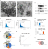
Biological characteristics of EVs from 3D cultured RM cells (3D-RM-EVs). (A) Transmission electron microscopy examination of 3D-RM-EVs and 2D-RM-EVs, bar 500 nm. (B) Western blot analysis of extracellular (CD63, CD81, and TSG101) and cellular (Calnexin) biomarkers of 3D-RM-EVs and 2D-RM-EVs. (C) Particle size analysis of 3D-RM-EVs and 2D-RM-EVs using nanoflow cytometry. (D) Comparison of the number of extracellular vesicles harvested from 3D cultural system with the amount harvested from conventional 2D culture. The error bars: standard errors of the mean from three independent experiments. (E) Comparison of the miRNA profiles of extracellular vesicles harvested from 3D cultural system with that from conventional 2D culture. (F) Correlation analysis of miRNA expression levels between 3D-RM-EVs and 2D-RM-EVs. (G) Shared miRNAs of 3D-RM-EVs and 2D-RM-EVs. EV: extracellular vesicle; RM: reserve mesenchymal. *P < 0.05; **P < 0.01.
Figure 4.
Figure 4.
3D-RM-EVs equipped with drugs inhibit mouse osteosarcoma cells proliferation in vitro. (A) Mouse osteosarcoma cells (K7M2 cells). (B) 3D-RM-EVs inhibited the proliferation of K7M2 cells, with cell viability decreased to 75% of normal level at 50 μg/ml of 3D-RM-EVs. The error bars: standard errors of the mean from three independent experiments. *P < 0.05; **P < 0.01. (C) Extracellular vesicles derived from human umbilical cord mesenchymal stem cells promote the proliferation of K7M2 cells. The error bars: standard errors of the mean from three independent experiments. *P < 0.05; **P < 0.01. (D) 3D-RM-EVs promoted the apoptosis of K7M2 cells, but the effect was not significant (P > 0.05). (E) Co-incubation mixture of 3D-RM-EVs and osteosarcoma therapeutic drugs (Ifosfamide + Etoposide) significantly inhibited K7M2 cells compared with positive drug group, *P < 0.05 at 1 μg/ml and/or more than 1 μg/ml. RM: reserve mesenchymal; EV: extracellular vesicle; UC-MSC: umbilical cord mesenchymal stem cell; IFO-ET: Ifosfamide + Etoposide; PE-A: Phycoerythrin A.
Figure 5.
Figure 5.
3D-RM-EVs equipped with drugs inhibit mouse osteosarcoma progression in vivo. (A) osteosarcoma mice (K7M2 bearing mice), histological analysis showed that the K7M2 osteosarcoma was a cancerous tissue attached to the periosteum of the bone, control: means normal tissue. (B) Tumor morphology observation showed that the 3D-RM-EVs + IFO + ET group had the most significant tumor inhibition effect, while the control group had the largest tumor. (C) Tumor weight analysis indicated that the tumor weight in the 3D-RM-EVs + IFO + ET group (2.12 ± 0.265 g) was significantly lighter than that in the control group (3.86 ± 0.232 g), 3D-RM-EVs group (3.70 ± 0.286 g), and IFO + ET group (2.92 ± 0.317 g). (D) Tumor growth curve analysis indicated that co-incubation of 3D-RM-EVs with osteosarcoma therapeutic drugs (Ifosfamide + Etoposide) had the best effect. (E) Weight changes of mice during treatment, there was no difference in weight between the 3D-RM-EVs group and the control group. The IFO + ET group had the most significant weight loss, while the 3D-RM-EVs + IFO + ET group could slow down weight loss compared with IFO + ET group. The error bars: standard errors of the mean from three independent experiments. RM: reserve mesenchymal; EV: extracellular vesicle; IFO-ET: Ifosfamide + Etoposide. *P < 0.05; **P < 0.01.

Similar articles

References

    1. Kourembanas S. Exosomes: vehicles of intercellular signaling, biomarkers, and vectors of cell therapy. Annu Rev Physiol. 2015;77:13–27. - PubMed
    1. Hassanzadeh A, Rahman HS, Markov A, Endjun JJ, Zekiy AO, Chartrand MS, Beheshtkhoo N, Kouhbanani M, Marofi F, Nikoo M, Jarahian M. Mesenchymal stem/stromal cell-derived exosomes in regenerative medicine and cancer; overview of development, challenges, and opportunities. Stem Cell Res Ther. 2021;12(1):297. - PMC - PubMed
    1. Chen BY, Sung CW, Chen C, Cheng CM, Lin DP, Huang CT, Hsu MY. Advances in exosomes technology. Clin Chim Acta. 2019;493:14–19. - PubMed
    1. Jin J, Shi Y, Gong J, Zhao L, Li Y, He Q, Huang H. Exosome secreted from adipose-derived stem cells attenuates diabetic nephropathy by promoting autophagy flux and inhibiting apoptosis in podocyte. Stem Cell Res Ther. 2019;10(1):95. - PMC - PubMed
    1. Qin Y, Wang L, Gao Z, Chen G, Zhang C. Bone marrow stromal/stem cell-derived extracellular vesicles regulate osteoblast activity and differentiation in vitro and promote bone regeneration in vivo. Sci Rep. 2016;6:21961. - PMC - PubMed

Publication types