. 2024 Feb;168(2):100-114.
doi: 10.1111/jnc.16029. Epub 2023 Dec 16.

Mechanisms of aquaporin-4 vesicular trafficking in mammalian cells

Affiliations

Mechanisms of aquaporin-4 vesicular trafficking in mammalian cells

Andrea Markou et al. J Neurochem. 2024 Feb.

Abstract

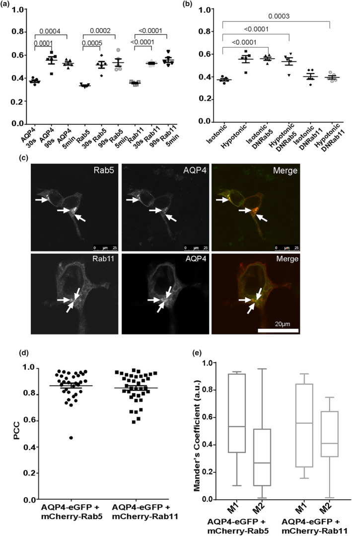
The aquaporin-4 (AQP4) water channel is abundantly expressed in the glial cells of the central nervous system and facilitates brain swelling following diverse insults, such as traumatic injury or stroke. Lack of specific and therapeutic AQP4 inhibitors highlights the need to explore alternative routes to control the water permeability of glial cell membranes. The cell surface abundance of AQP4 in mammalian cells fluctuates rapidly in response to changes in oxygen levels and tonicity, suggesting a role for vesicular trafficking in its translocation to and from the cell surface. However, the molecular mechanisms of AQP4 trafficking are not fully elucidated. In this work, early and recycling endosomes were investigated as likely candidates of rapid AQP4 translocation together with changes in cytoskeletal dynamics. In transiently transfected HEK293 cells a significant amount of AQP-eGFP colocalised with mCherry-Rab5-positive early endosomes and mCherry-Rab11-positive recycling endosomes. When exposed to hypotonic conditions, AQP4-eGFP rapidly translocated from intracellular vesicles to the cell surface. Co-expression of dominant negative forms of the mCherry-Rab5 and -Rab11 with AQP4-eGFP prevented hypotonicity-induced AQP4-eGFP trafficking and led to concentration at the cell surface or intracellular vesicles respectively. Use of endocytosis inhibiting drugs indicated that AQP4 internalisation was dynamin-dependent. Cytoskeleton dynamics-modifying drugs also affected AQP4 translocation to and from the cell surface. AQP4 trafficking mechanisms were validated in primary human astrocytes, which express high levels of endogenous AQP4. The results highlight the role of early and recycling endosomes and cytoskeletal dynamics in AQP4 translocation in response to hypotonic and hypoxic stress and suggest continuous cycling of AQP4 between intracellular vesicles and the cell surface under physiological conditions.

Keywords: Rab GTPase; aquaporin-4; astrocyte; cytoskeleton; oedema; vesicular trafficking.


Conflict of interest statement

R.M.B., P.K. and M.M.S. are shareholders in Estuar Pharmaceuticals.

Figures

FIGURE 1
FIGURE 1
AQP4‐eGFP translocation in HEK293 cells following dynasore and filipin treatment. HEK293 cells were transfected with AQP4‐eGFP and incubated in the presence of filipin and dynasore as described in the methods. AQP4‐eGFP localisation was imaged by live fluorescence microscopy in an isotonic solution at 30 s, at 5 min following exposure to a hypotonic medium, and at 5 min following return back to isotonic medium. (a) representative schematic of how HEK293 transfected with AQP4‐eGFP were analysed to determine relative membrane expression (RME). (b) RME quantification of AQP4‐eGFP in control and dynasore‐treated cells. (c) RME quantification of AQP4‐eGFP in control and filipin‐treated cells. Mean RME for AQP4‐eGFP averaged over three profiles/cell of at least 10 cells per condition in each repeat in three experimental repeats, n = 3. For each repeat, p values are from one‐way analysis of variance followed by Bonferroni's correction. All data are presented as mean ± SEM.
FIGURE 2
FIGURE 2
AQP4‐eGFP translocation in HEK293 cells in the presence of cytoskeleton‐modifying drugs. HEK293 cells were transfected with AQP4‐eGFP and incubated in the presence of cytoskeleton modifying drugs as described in the Methods. AQP4‐eGFP localisation was imaged by live fluorescence microscopy in an isotonic solution at 30 s, at 5 min following exposure to a hypotonic medium, and at 5 min following return back to isotonic medium. Quantification of AQP‐eGFP relative membrane expression (RME) in AQP4 + nocodazole (a), AQP4 + cytochalasin D (b), AQP4 + paclitaxel (c) AQP4 + jasplakinolide (d). Mean RME for AQP4‐eGFP averaged over three profiles/cell of at least 10 cells per condition in each repeat in three experimental repeats, n = 3. For each repeat, p values are from one‐way analysis of variance followed by Bonferroni's correction. All data are presented as mean ± SEM.
FIGURE 3
FIGURE 3
AQP4‐eGFP translocation in HEK293 cells via early and recycling endosomes. HEK293 cells were transfected with AQP4‐eGFP in combination with one of the following: mCherry‐Rab5, mCherry‐Rab11, mCherry‐Rab5DN or DsRed‐Rab11DN. AQP4‐eGFP localisation was imaged by live fluorescence microscopy in an isotonic solution at 30 s, at 5 min following exposure to a hypotonic medium and at 5 min following return back to isotonic medium. Mean RME for AQP4‐eGFP and mCherry‐tagged Rab5 and Rab11 (a), and AQP4eGFP co‐transfected with Rab5DN and Rab11DN (b), averaged over three profiles/cell of at least 10 cells per condition in each repeat in three experimental repeats, n = 3. Cells transiently transfected with AQP4‐eGFP (green), mCherry‐Rab5 or mCherry‐Rab11 (red) and the merged images (yellow) (c). Images of each channel were taken sequentially at 63x magnification. The localisation of AQP4‐eGFP and mCherry‐Rab5 overlaps in many areas (white arrows), as does the localisation of AQP4‐eGFP and mCherry‐Rab11 (white arrows). The scale bar is 25 μm. Correlation (PCC) in selected cells (d) and co‐occurrence (e) in selected cells of AQP4‐eGFP with mCherry‐Rab5 and colocalisation of AQP4‐eGP with mCherry‐Rab11. For each repeat, p values are from a one‐way analysis of variance followed by Bonferroni's correction. All data are presented as mean ± SEM.
FIGURE 4
FIGURE 4
AQP4 internalisation in primary human astrocytes following dynasore and filipin treatment. Surface expression of endogenous AQP4 in primary human astrocytes following treatments was measured by surface biotinylation assay as described in the methods. Treatment with DMSO was used as a vesicle control. (a) Surface expression of AQP4 in control and dynasore‐treated cells in hypotonic and isotonic conditions; (b) surface expression of AQP4 in control and filipin‐treated cells in hypotonic and isotonic conditions; (c) (d) percentage of internalised AQP4 in isotonic and hypotonic media following dynasore and filipin treatment respectively. (e) Surface expression of AQP4 in control, hypoxic and hypotonic conditions following treatment with dynasore and filipin. Control black bar represents normoxic and isotonic conditions; n = 3 for each repeat, p‐values are from one‐way analysis of variance followed by Tukey's correction. All data are presented as mean ± SEM.
FIGURE 5
FIGURE 5
AQP4 translocation in primary human astrocytes in response to hypoxia and hypotonicity following treatment with cytoskeleton‐modifying drugs. Surface expression of endogenous AQP4 in primary human astrocytes following exposure to hypotonic medium and return back to isotonic medium was measured by surface biotinylation assay as described in the methods following pre‐incubation with paclitaxel (a), nocodazole (b), jasplakinolide (c) and cytochalasin D (d). Treatment with DMSO was used as a vesicle control. (e–h) percentage of internalised AQP4 following exposure to hypotonic medium and pre‐treatments with paclitaxel, nocodazole, jasplakinolide and cytochalasin D respectively. I, surface expression of endogenous AQP4 in primary human astrocytes following exposure to hypotonic medium or hypoxia measured by surface biotinylation assay n = 3 for each repeat, p‐values are from one‐way analysis of variance followed by Tukey's correction. All data are presented as mean ± SEM.
FIGURE 6
FIGURE 6
Molecular mechanisms of AQP4 translocation in mammalian cells. AQP4 continuously cycles between the cell surface, Rab5‐positive early and Rab11‐positive recycling endosomes in mammalian cells. AQP4 internalisation is dynamin‐dependent and AQP4 translocation mechanisms are impaired upon inhibition of Rab5 and Rab11 function as well as cytoskeleton dynamics using cytoskeleton dynamics‐modifying drugs. Created with biorender.com.

References

    1. Aoki, T. , Suzuki, T. , Hagiwara, H. , Kuwahara, M. , Sasaki, S. , Takata, K. , & Matsuzaki, T. (2012). Close association of aquaporin‐2 internalization with caveolin‐1. Acta Histochemica et Cytochemica, 45(2), 139–146. - PMC - PubMed
    1. Badaut, J. , Lasbennes, F. , Magistretti, P. J. , & Regli, L. (2002). Aquaporins in brain: Distribution, physiology, and pathophysiology. Journal of Cerebral Blood Flow and Metabolism, 22(4), 367–378. - PubMed
    1. Bandmann, V. , Mirsanaye, A. S. , Schäfer, J. , Thiel, G. , Holstein, T. , & Mikosch‐Wersching, M. (2019). Membrane capacitance recordings resolve dynamics and complexity of receptor‐mediated endocytosis in Wnt signalling. Scientific Reports, 9(1), 12999. - PMC - PubMed
    1. Carmosino, M. , Procino, G. , Tamma, G. , Mannucci, R. , Svelto, M. , & Valenti, G. (2007). Trafficking and phosphorylation dynamics of AQP4 in histamine‐treated human gastric cells. Biology of the Cell, 99(1), 25–36. - PubMed
    1. Casella, J. F. , Flanagan, M. D. , & Lin, S. (1981). Cytochalasin D inhibits Actin polymerization and induces depolymerization of Actin filaments formed during platelet shape change. Nature, 293(5830), 302–305. - PubMed

Publication types