

doi: 10.4103/1673-5374.389644.
Epub 2023 Dec 11.
Suppression of mature TAU isoforms prevents Alzheimer's disease-like amyloid-beta oligomer-induced spine loss in rodent neurons
Affiliations
- PMID: 38103227
- PMCID: PMC10960270
- DOI: 10.4103/1673-5374.389644
Item in Clipboard
Suppression of mature TAU isoforms prevents Alzheimer's disease-like amyloid-beta oligomer-induced spine loss in rodent neurons
Neural Regen Res.
.
No abstract available
Figures
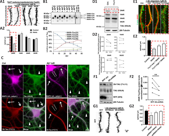
2N4R-TAU is necessary and sufficient for AβO-induced spine loss in primary neurons. (A) Reintroduction of different TAU isoforms into primary murine TAU-KO neurons after the establishment of neuronal cell polarity and spine formation (days in vitro (DIV) 15). Different isoforms of human Dendra2c-tagged TAU (TAUD2) were co-transfected with tdTomato (as volume marker) for five days, afterwards, cells were treated with 1 µM AβO for 3 hours. (A1) Fluorescence microscopy of the volume marker tdTomato shows a reduction of dendritic spines after transfection with 2N4R-TAU followed by AβO treatment, but not in other conditions. Scale bar: 4 µm. (A2) Quantification of A1. n = 5–10 dendrites, at least 5 cells were analyzed per condition. (B) Time course of endogenous TAU isoform expression of rat primary neurons between DIV2 and DIV49. (B1) Western blot of rat cortical neurons grown for the indicated time points. Samples were left untreated (-AP) or treated with alkaline phosphatase (+AP) for dephosphorylation. (B2) Quantification of B1. Only after 3 weeks of culture, the longest rodent TAU isoform (2N4R-TAU) is detectable, while the shortest rodent TAU isoform (0N3R-TAU) steadily decreases until the end of the analysis. Note that until three weeks in vitro, 0N3R-TAU constitutes the majority of the TAU protein in cultured neurons. n = 3–4 independent cultures. (C) Immunofluorescence images of mature primary rat neurons (DIV35) after vehicle control and AβO treatment. Neurons were stained for 2N-TAU (71C11), the dendritic marker MAP2, and an antibody recognizing all TAU isoforms (K9JA). Neurons express solid levels of 2N4R-TAU in the soma and dendrites (indicated by arrows) but not in the axons (indicated by arrowheads). The dotted lines indicate axon hillock. Treatment with AβO results in an increased signal of 2N-TAU in the dendrites. (D–E) 2N4R-TAU can be suppressed by 2N-TAU specific antibody (71C11, incubation for 4–5 days) in rat cortical neurons, which prevents AβO-induced spine loss. (D1) Western blot with a panTAU antibody (K9JA) of dephosphorylated TAU of mature rat primary neurons (35 DIV) shows that 2N4R-TAU is decreased after exposure to 71C11 (1:600 dilution) for 4–5 days. βIII-tubulin was used as a loading control. (D2) Quantification of individual TAU isoform expression in D1. n = 3 independent cultures. (E1) (Immuno-) Fluorescence images of mature primary neurons (DIV35) incubated with 71C11 as indicated. Neurons were treated with either vehicle control or AβO for 3 hours and afterwards stained for F-actin as an indicator for dendritic spines and MAP2 as a dendrite marker. While neurons treated with AβO show pronounced loss of spines, this is largely prevented by pre-incubation of cells with 71C11 (red dotted box). (E2) Quantification of spine density after knockdown of 2N-TAU by 71C11 and treatment with vehicle control or 1 µM AβO. n = 5–10 dendrites of different cells. (F–G) AdV-based shRNA-mediated knockdown of 2N4R-TAU prevents AβO-induces spine loss in rat primary neurons. (F1) Western blot with 2N-TAU specific (71C11) and panTAU antibody (K9JA) of dephosphorylated TAU of mature primary neurons (35 DIV) after transduction with AdV-RFP or AdV-2N-TAU shRNA for 3–4 days. 2N4R-TAU expression is decreased after AdV-mediated KD. βIII-tubulin was used as a loading control. (F2) Quantification of F1. Expression in primary rat neurons after transduction with AdV-2N-TAU shRNA. n = 4 independent cultures. (G1) Immunofluorescence images of mature primary neurons (DIV35) after AdV-transduction and AβO treatment as indicated. Transduced cells express RFP, which is used as a volume marker to visualize spines. While control neurons treated with AβO show pronounced loss of spines, this is largely prevented by KD of 2N4R-TAU. (G2) Quantification of G1. Spine density after KD of 2N-TAU by shRNA and treatment with vehicle control or 1 µM AβO. n = 5–10 dendrites of different cells. Error bars represent SEM. Statistical analysis was performed by t-test with Tukey's test for correction of multiple comparisons. *P < 0.05; **P < 0.01; ***P < 0.001. Arrows indicate dendrites; arrowheads indicate axons; red dotted lines highlight important findings; white dotted lines indicate axon hillocks. Paired analysis was done to compare the treated vs. untreated cultures, due to high differences between individual cultures. Unpublished data by our team. AdV: Adenovirus; AP: alkaline phosphatase; AβO: amyloid beta oligomers; DIV: days in vitro; KD: knockdown; TAUD2: Dendra2c-tagged TAU.
References
-
- Buchholz S, Bell-Simons M, Kabbani MAA, Kluge L, Cagkmak C, Klimek J, Zempel H. The TAU isoform 1N4R restores vulnerability of MAPT knockout human iPSC-derived neurons to amyloid beta-induced neuronal dysfunction. Res Sq. 2022 doi:10.21203/rs.3.rs-2277268/v1.
-
- Bullmann T, Holzer M, Mori H, Arendt T. Pattern of tau isoforms expression during development in vivo. Int J Dev Neurosci. 2009;27:591–597. - PubMed
-
- Goedert M, Spillantini MG, Jakes R, Rutherford D, Crowther RA. Multiple isoforms of human microtubule-associated protein tau: sequences and localization in neurofibrillary tangles of Alzheimer's disease. Neuron. 1989;3:519–526. - PubMed
LinkOut - more resources
Full Text Sources
