

Contributions of diverse models of the female reproductive tract to the study of Chlamydia trachomatis-host interactions
- PMID: 38103413
- PMCID: PMC10922760
- DOI: 10.1016/j.mib.2023.102416
Contributions of diverse models of the female reproductive tract to the study of Chlamydia trachomatis-host interactions
Abstract
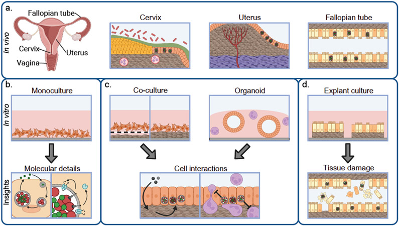
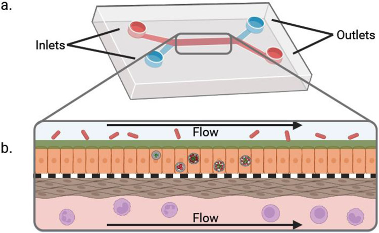
Chlamydia trachomatis is a common cause of sexually transmitted infections in humans with devastating sequelae. Understanding of disease on all scales, from molecular details to the immunology underlying pathology, is essential for identifying new ways of preventing and treating chlamydia. Infection models of various complexity are essential to understand all aspects of chlamydia pathogenesis. Cell culture systems allow for research into molecular details of infection, including characterization of the unique biphasic Chlamydia developmental cycle and the role of type-III-secreted effectors in modifying the host environment to allow for infection. Multicell type and organoid culture provide means to investigate how cells other than the infected cells contribute to the control of infection. Emerging comprehensive three-dimensional biomimetic systems may fill an important gap in current models to provide information on complex phenotypes that cannot be modeled in simpler in vitro models.
Copyright © 2023 Elsevier Ltd. All rights reserved.
Conflict of interest statement
Declaration of Competing Interest The authors declare no competing interests relevant to this article.
Figures
Similar articles
-
Murine Endometrial Organoids to Model Chlamydia Infection.Front Cell Infect Microbiol. 2020 Aug 14;10:416. doi: 10.3389/fcimb.2020.00416. eCollection 2020. Front Cell Infect Microbiol. 2020. PMID: 32923409 Free PMC article.
-
Early Transcriptional Landscapes of Chlamydia trachomatis-Infected Epithelial Cells at Single Cell Resolution.Front Cell Infect Microbiol. 2019 Nov 19;9:392. doi: 10.3389/fcimb.2019.00392. eCollection 2019. Front Cell Infect Microbiol. 2019. PMID: 31803632 Free PMC article.
-
Activation of neutrophils by Chlamydia trachomatis-infected epithelial cells is modulated by the chlamydial plasmid.Microbes Infect. 2018 May;20(5):284-292. doi: 10.1016/j.micinf.2018.02.007. Epub 2018 Mar 2. Microbes Infect. 2018. PMID: 29499390
-
Immunology of Chlamydia infection: implications for a Chlamydia trachomatis vaccine.Nat Rev Immunol. 2005 Feb;5(2):149-61. doi: 10.1038/nri1551. Nat Rev Immunol. 2005. PMID: 15688042 Review.
-
The mucosal immune response to Chlamydia trachomatis infection of the reproductive tract in women.J Reprod Immunol. 2009 Dec;83(1-2):173-8. doi: 10.1016/j.jri.2009.07.013. Epub 2009 Nov 5. J Reprod Immunol. 2009. PMID: 19896206 Review.
Cited by
-
Study Models for Chlamydia trachomatis Infection of the Female Reproductive Tract.Microorganisms. 2025 Feb 28;13(3):553. doi: 10.3390/microorganisms13030553. Microorganisms. 2025. PMID: 40142446 Free PMC article. Review.
References
-
- Haggerty CL, Gottlieb SL, Taylor BD, Low N, Xu F, Ness RB: Risk of sequelae after Chlamydia trachomatis genital infection in women. J Infect Dis 2010, 201 Suppl 2:S134–155. - PubMed
-
- Karim S, Souho T, Benlemlih M, Bennani B: Cervical Cancer Induction Enhancement Potential of Chlamydia trachomatis: A Systematic Review. Curr Microbiol 2018, 75:1667–1674. - PubMed
-
- Abdelrahman YM, Belland RJ: The chlamydial developmental cycle. FEMS Microbiol Rev 2005, 29:949–959. - PubMed
Publication types
MeSH terms
Grants and funding
LinkOut - more resources
Full Text Sources
Medical
Miscellaneous
