. 2024 Feb;39(1):97-112.
doi: 10.1016/j.virs.2023.12.003. Epub 2023 Dec 14.

Phosphorylation of PB2 at serine 181 restricts viral replication and virulence of the highly pathogenic H5N1 avian influenza virus in mice

Affiliations

Phosphorylation of PB2 at serine 181 restricts viral replication and virulence of the highly pathogenic H5N1 avian influenza virus in mice

Jiao Hu et al. Virol Sin. 2024 Feb.

Abstract

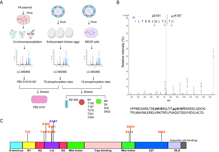
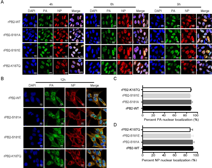
Influenza A virus (IAV) continues to pose a pandemic threat to public health, resulting a high mortality rate annually and during pandemic years. Posttranslational modification of viral protein plays a substantial role in regulating IAV infection. Here, based on immunoprecipitation (IP)-based mass spectrometry (MS) and purified virus-coupled MS, a total of 89 phosphorylation sites distributed among 10 encoded viral proteins of IAV were identified, including 60 novel phosphorylation sites. Additionally, for the first time, we provide evidence that PB2 can also be acetylated at site K187. Notably, the PB2 S181 phosphorylation site was consistently identified in both IP-based MS and purified virus-based MS. Both S181 and K187 are exposed on the surface of the PB2 protein and are highly conserved in various IAV strains, suggesting their fundamental importance in the IAV life cycle. Bioinformatic analysis results demonstrated that S181E/A and K187Q/R mimic mutations do not significantly alter the PB2 protein structure. While continuous phosphorylation mimicked by the PB2 S181E mutation substantially decreases viral fitness in mice, PB2 K187Q mimetic acetylation slightly enhances viral virulence in mice. Mechanistically, PB2 S181E substantially impairs viral polymerase activity and viral replication, remarkably dampens protein stability and nuclear accumulation of PB2, and significantly weakens IAV-induced inflammatory responses. Therefore, our study further enriches the database of phosphorylation and acetylation sites of influenza viral proteins, laying a foundation for subsequent mechanistic studies. Meanwhile, the unraveled antiviral effect of PB2 S181E mimetic phosphorylation may provide a new target for the subsequent study of antiviral drugs.

Keywords: Acetylation; H5N1 influenza virus; Mice; PB2; Phosphorylation; Viral fitness.


Figures

Fig. 1
Fig. 1
Identification of the PB2 S181 phosphorylation site and K187 acetylation site using IP-based MS and purified virus-coupled MS. A The flow chart for identification of phosphorylation and acetylation sites that located in the H5N1 virus CK10 strain. For the IP samples, a total of 3 ​× ​106 HEK 293T cells were transfected with 12 ​μg of pcDNA-PA plasmid. After 24 ​h, cells were infected with 2 MOI of the CK10 virus. After 8 ​h, cells were lysed for preparation of IP sample using PA as the primary antibody. In addition, MS was also performed on the purified viral samples inoculated in MDCK cells and embryonated chicken eggs. For MDCK cells, cells were infected with CK10 virus at an MOI of 4. B MS analysis results revealed the phosphorylation of PB2 S181 and acetylation of PB2 K187. “P” means the phosphorylation site, while “AC” stands for the acetylation site. C Function analysis of the PB2 phosphorylation sites and PB2 acetylation site K187.
Fig. 2
Fig. 2
PB2 S181 and PB2 K187 are exposed on the surface of the PB2 protein. A The surface accessibility analysis of PB2 S181 and K187 using NetSurfP 3.0. The first line plot shows the amino acid letter. The relative surface area (RSA) is shown by a filled curve, threshold at 25% (red exposed, blue buried). The secondary structure is as annotated as Q3 and shows the symbols as a-helix (screw), beta-sheet (arrows), coil (line). The disorder is a grey line in which its thickness is equal to the probability of it being a disordered region. Reported accuracy on test datasets for predicted protein features: relative surface area (RSA), accessible surface area (ASA), secondary structure (SS8 and SS3), protein disorder, and Phi and Psi torsion angles. B The overall 3D-structure analysis of the PB2 protein by SWISS-MODEL (SWISS-MODEL.expasy.org) using the PyMOL software. The position of S181 in PB2 was shown in green, the location of K187 in PB2 was shown in blue. C Enlarged 3D-structure of the PB2 protein indicating the specific location of S181 and K187 in PB2. DF Determine the position of PB2 S181 and K187 in the assembled polymerase complex. The crystal structures of the polymerase were predicted using SWISS-MODEL Homology Modeling (ProMod3 3.3.0). The PDB: 8h69.1 Cryo-EM structure of influenza RNA polymerase was served as the template.
Fig. 3
Fig. 3
The root mean square deviation (RMSD) analysis between the wild-type PB2 and the mutant PB2 protein. The wild-type and the mutant protein sequences were uploaded into the RoseTTAFold server for protein structure prediction. The predicted structures of the wild-type and the mutant proteins were then downloaded into PyMOL software for alignment analysis. The RMSD <1, means PB2 mimic-modification mutations do not significantly alter the structure of the PB2 protein. A Alignment of the PB2-WT and the mutant PB2-S181A protein. B Alignment of the PB2-WT and the mutant PB2-S181E protein. C Color gradation for each PB2 protein. D Alignment of the PB2-WT and the mutant PB2-K187Q protein. E Alignment of the PB2-WT and the mutant PB2-S181R protein.
Fig. 4
Fig. 4
Effect of the PB2-S181E/A and PB2-S187Q/R mimic mutation on PB2 protein stability. Human HEK 293T cells were transfected with 1 ​μg of wild-type or mutant pcDNA3.1-PB2 expression plasmid. After 24 ​h, 100 ​μg/mL of the CHX was added to each dish. After 0 ​h of CHX treatment, samples were collected at 1 ​h intervals thereafter until 4 ​h. Samples were then collected for Western bolting analysis. A Western bolting results of the PB2 protein expression after treat with the CHX. B Relative intensity of the PB2-WT and PB2-S181 ​E/A mutant protein from panel A. C Relative intensity of the PB2-WT and PB2-S187Q/R mutant protein from panel A. For 4B and 4C, data was shown as the mean ​± ​SD of three independent experiments. For each replicate, PB2 protein levels were normalized to tubulin protein levels.
Fig. 5
Fig. 5
Impact of PB2-S181E/A and PB2-S187Q/R mimic mutation on viral polymerase activity. AB Effect of PB2-S181E/A (A) and PB2-S187Q/R (B) mimic mutation on viral polymerase activities by mini-genome assay. 293T cells were transfected in triplicate with luciferase reporter plasmid p-Luci and internal control plasmid Renilla pRL-TK, together with plasmids expressing PB2, PB1, PA, and NP from CK10 virus or the indicated PB2 mutants. At 24 ​h p.t., cell lysates were used to measure firefly and Renilla luciferase activities. Values are shown as the means ​± ​SD of the three independent experiments and are standardized to those of PB2-WT (100%). C Determine the expression of viral proteins in panel A by WB. D Determine the expression of viral proteins in panel B by WB. E Calculating the relative expression of the viral proteins in panel C. F Calculation of the relative expression of the viral proteins in panel D. Values in panel E and F are shown as the means ​± ​SD of the three independent experiments and are standardized to those of PB2-WT (1.0).
Fig. 6
Fig. 6
Impact of PB2-S181E/A and PB2-S187Q/R mimic mutation on viral replication in vitro. Cells were inoculated at an MOI of 0.01. Data was shown as the mean ​± ​SD of three independent infections. AB Effect of PB2-S181E/A and PB2-S187Q mimic mutation on viral replication in MDCK cells (A) and DF-1 ​cells (B). Virus titers were determined as TCID50 in MDCK cells at the indicated time points. CD Effect of PB2-S181E/A and PB2-S187Q mimic mutation on NP mRNA/cRNA/vRNA expression in MDCK cells (C), and DF-1 ​cells (D). EF Effect of PB2-S181E/A and PB2-S187Q mimic mutation on NP and PB2 protein expression in MDCK cells (E), and DF-1 ​cells (F). G Calculatiion of the relative expression of the viral proteins in panel E. H Calculating the relative expression of the viral proteins in panel F.
Fig. 7
Fig. 7
Influence of PB2-S181E/A and PB2-S187Q mimic mutation on PB2 nuclear accumulation in MDCK and DF1 cells. MDCK or DF1 cells were infected with the indicated virus at an MOI of 2, cell cultures were then fixed and processed for immunofluorescence observation at indicated time points. Cell nuclei were stained with DAPI. A At 4 ​h, 6 ​h, 9 ​h and 12 ​h p.i., MDCK cells were fixed and processed for immunofluorescence observation. Scale bar, 14 μm. B At 4 ​h p.i., MDCK cells were fixed and processed for immunofluorescence observation. Scale bar, 6 μm. C At 4 ​h p.i., the PB2 nuclear accumulation in MDCK-infected cells was determined as the ratio of cells showing green fluorescence in the nucleus to the total number of cells counted (n ​= ​200). D At 4 ​h, 6 ​h, 9 ​h, and 12 h p.i., DF1 cells were fixed and processed for immunofluorescence observation. Scale bar, 16 μm. E At 4 ​h p.i., the PB2 nuclear accumulation in DF1-infected cells was determined as the ratio of cells showing green fluorescence in the nucleus to the total number of cells counted (n ​= ​200). Values shown are the means of the results of three independent experiments SDs. ∗, P ​< ​0.05 compared with the result for rPB2-WT virus-infected cells.
Fig. 8
Fig. 8
Effect of PB2-S181E/A and PB2-S187Q mimic mutation on viral virulence in mice. A Body weight change of the infected mice challenged with 105.0 EID50 of the CK10 virus. Body weight was presented as percentage of the weight on the day of inoculation (day 0). Mice were humanely killed when they lost >25% of their initial body weight. B Survival rate of the infected mice. Mice were infected with the indicated virus and were observed for 14 days. C MLD50 value of the recombinant and parental viruses. D Histopathology in the mouse lung-infected with the indicated virus in a dose of 105.0 EID50.☆, lung congestion and hemorrhage; □, necrotic detached cells were seen in the bronchiolar lumen; , lymphocyte infiltration in pulmonary alveoli bronchiolar lumen and around blood vessel; formula image, pulmonary edema. E Scores of the histopathological changes in the mouse lung on day 3 and 5 p.i..
Fig. 9
Fig. 9
Viral replication in mouse organs. Groups of mice were infected with the indicated recombinant virus. A-B Three mice of each group that infected with 105.0 EID50 of the viruses were euthanized on day 3 and 5 p.i. for determination of viral load in mice organs; C Mouse lung was collected for detection of the NP antigen expression using IFA. Scale bar, 75 μm. D-E The immunofluorescent intensity of the NP antigen from panel C was calculated, day 3 p.i. (D) and day 5 p.i. (E).
Fig. 10
Fig. 10
Impact of the PB2-S181E/A and PB2-S187Q mimic mutation on innate immune response in mouse lung. Groups of mice were infected with the indicated recombinant virus at a dose of 105.0 EID50. Three mice of each group were euthanized on day 3 and 5 p.i. for determination of cytokine responses in mouse lung. The expression of cytokines in mouse lung was analyzed by qRT-PCR. Values were shown as the mean ​± ​SD of three samples.
figs1
figs1
figs2
figs2
figs3
figs3

Similar articles

References

    1. Agüero M., Monne I., Sánchez A., Zecchin B., Fusaro A., Ruano M.J., Del Valle Arrojo M., Fernández-Antonio R., Souto A.M., Tordable P., Cañás J., Bonfante F., Giussani E., Terregino C., Orejas J.J. Highly pathogenic avian influenza A(H5N1) virus infection in farmed minks, Spain, October 2022. Euro Surveill. 2023;28 - PMC - PubMed
    1. Boergeling Y., Brunotte L., Ludwig S. Dynamic phospho-modification of viral proteins as a crucial regulatory layer of influenza A virus replication and innate immune responses. Biol. Chem. 2021;402:1493–1504. - PubMed
    1. Cazals F., Tetley R. Characterizing molecular flexibility by combining least root mean square deviation measures. Proteins. 2019;87:380–389. - PubMed
    1. Chen H., Qian Y.J., Chen X., Ruan Z.Y., Ye Y.T., Chen H.J., Babiuk L.A., Jung Y.S., Dai J.J. HDAC6 restricts influenza A virus by deacetylation of the RNA polymerase PA subunit. J. Virol. 2019;93, e01896-18 - PMC - PubMed
    1. Chen L., Wang C.M., Luo J., Su W., Li M., Zhao N., Lyu W.T., Attaran H., He Y.P., Ding H., He H.X. Histone deacetylase 1 plays an acetylation-independent role in influenza A virus replication. Front. Immunol. 2017;8, 1757 - PMC - PubMed

Substances