

Sclerosing epithelioid fibrosarcoma associated with WRN gene variant presenting as chronic dyspnea and pathologic cervical fracture: a case report and review of the literature
- PMID: 38104125
- PMCID: PMC10725598
- DOI: 10.1186/s13256-023-04249-6
Sclerosing epithelioid fibrosarcoma associated with WRN gene variant presenting as chronic dyspnea and pathologic cervical fracture: a case report and review of the literature
Abstract
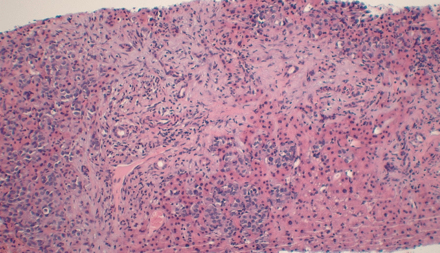
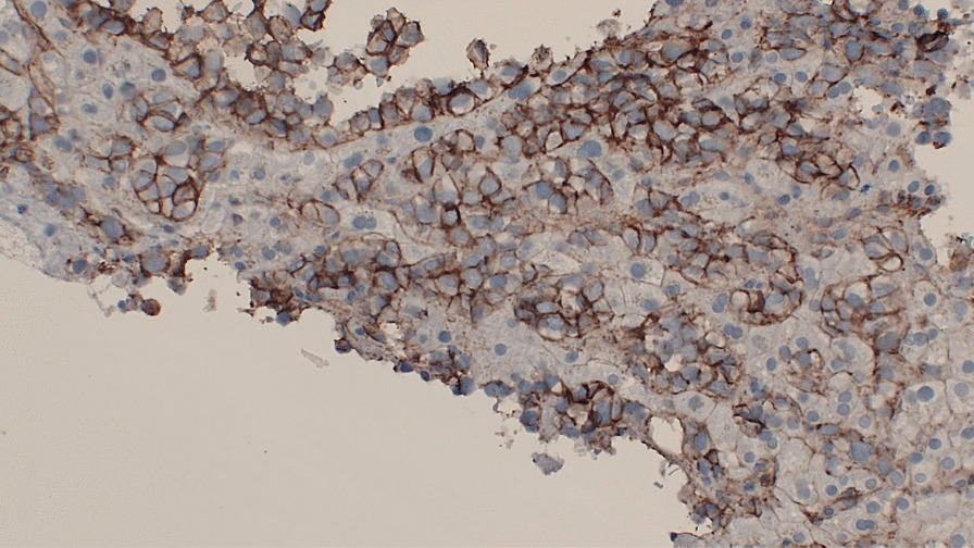
Background: Sclerosing epithelioid fibrosarcoma is an aggressive sarcoma subtype with poor prognosis and limited response to conventional chemotherapy regimens. Diagnosis can be difficult owing to its variable presentation, and cases of sclerosing epithelioid fibrosarcoma are rare. Sclerosing epithelioid fibrosarcoma typically affects middle-aged individuals, with studies inconsistently citing gender predominance. Sclerosing epithelioid fibrosarcoma typically arises from the bones and soft tissues and often has local recurrence after resection and late metastases. Immunohistochemical staining typically is positive for mucin-4. Werner syndrome is due to an autosomal recessive mutation in the WRN gene and predisposes patients to malignancy.
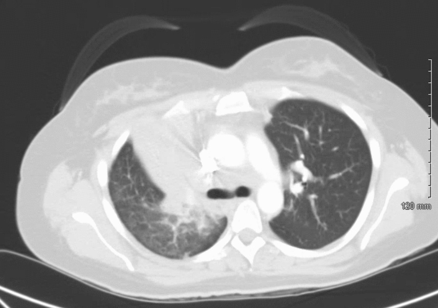
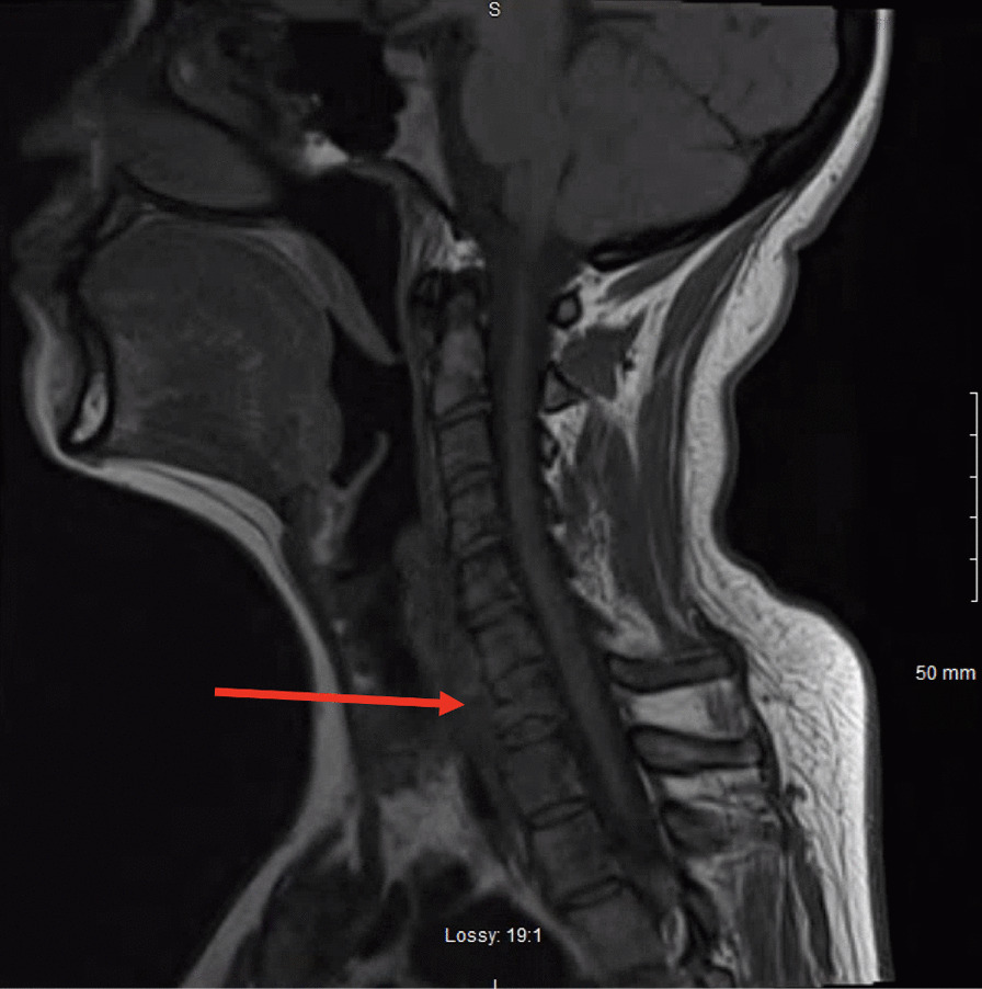
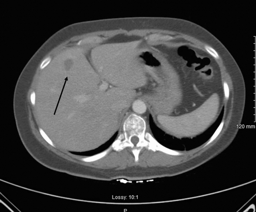
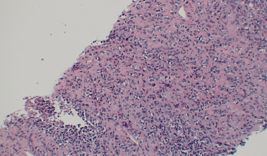
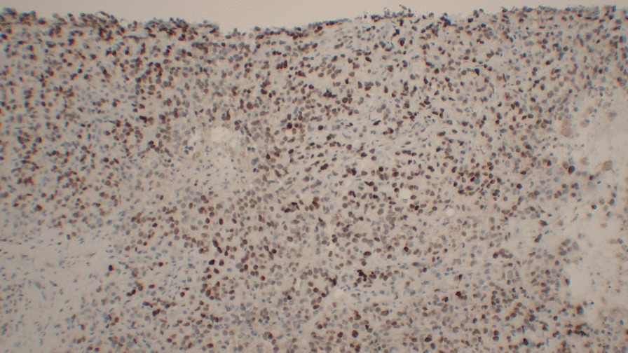
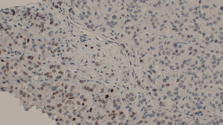
Case presentation: A 37-year-old Caucasian female presented to the emergency department with 4 months of dyspnea and back pain. She had been treated for pneumonia but had persistent symptoms. A chest, abdomen, and pelvis computed tomography showed near-complete right upper lobe collapse and consolidation, mediastinal lymphadenopathy, lytic spinal lesions, and a single 15-mm hypodense liver nodule. The patient underwent a transthoracic right upper lobe biopsy, bronchoscopy, endobronchial ultrasound with transbronchial lymph node sampling, and bronchoalveolar lavage of the right upper lobe. The bronchoalveolar lavage cytology was positive for malignant cells compatible with poorly differentiated non-small cell carcinoma; however, the cell block materials were insufficient to run immunostains for further investigation of the bronchoalveolar lavage results. Consequently, the patient also underwent a liver biopsy of the liver nodule, which later confirmed a diagnosis of sclerosing epithelioid fibrosarcoma. Next-generation sequencing revealed a variant of unknown significance in the WRN gene. She was subsequently started on doxorubicin.
Conclusion: Sclerosing epithelioid fibrosarcoma is a very rare entity, only cited approximately 100 times in literature to date. Physicians should be aware of this disease entity and consider it in their differential diagnosis. Though pulmonary involvement has been described in the context of sclerosing epithelioid fibrosarcoma, this malignancy may affect many organ systems, warranting extensive investigation. Through our diagnostic workup, we suggest a possible link between sclerosing epithelioid fibrosarcoma and the WRN gene. Further study is needed to advance our understanding of sclerosing epithelioid fibrosarcoma and its clinical associations as it is an exceedingly rare diagnosis.
Keywords: Bronchoscopy; Pathology; Pulmonology; Sclerosing epithelioid fibrosarcoma; Thoracic oncology.
© 2023. This is a U.S. Government work and not under copyright protection in the US; foreign copyright protection may apply.
Conflict of interest statement
The authors declare that they have no competing interests.
Figures
References
-
- Doyle LA, Wang WL, Dal Cin P, Lopez-Terrada D, Mertens F, Lazar AJ, Fletcher CD, Hornick JL. MUC4 is a sensitive and extremely useful marker for sclerosing epithelioid fibrosarcoma: association with FUS gene rearrangement. Am J Surg Pathol. 2012;36(10):1444–1451. doi: 10.1097/PAS.0b013e3182562bf8. - DOI - PubMed
Publication types
MeSH terms
Substances
LinkOut - more resources
Full Text Sources
Medical
