

Overexpress miR-132 in the Brain Parenchyma by a Non-invasive Way Improves Tissue Repairment and Releases Memory Impairment After Traumatic Brain Injury
- PMID: 38104297
- PMCID: PMC11397820
- DOI: 10.1007/s10571-023-01435-4
Overexpress miR-132 in the Brain Parenchyma by a Non-invasive Way Improves Tissue Repairment and Releases Memory Impairment After Traumatic Brain Injury
Abstract
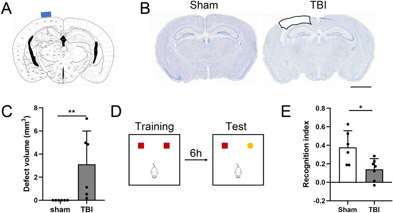
Traumatic brain injury (TBI) is a serious public health problem worldwide, which could lead to an extremely high percentage of mortality and disability. Current treatment strategies mainly concentrate on neuronal protection and reconstruction, among them, exogenous neural stem cell (NSC) transplantation has long been regarded as the most effective curative treatment. However, due to secondary trauma, transplant rejection, and increased incidence of brain malignant tumor, a non-invasive therapy that enhanced endogenous neurogenesis was more suitable for TBI treatment. Our previous work has shown that miR-132 overexpression could improve neuronal differentiation of NSCs in vitro and in vivo. So, we engineered a new kind of AAV vector named AAV-PHP.eB which can transfect brain parenchyma through intravenous injection to overexpress miR-132 in brain after TBI. We found that miR-132 overexpression could reduce impact volume, promote neurogenesis in the dentate gyrus (DG), accelerate neuroblast migrating into the impact cortex, ameliorate microglia-mediated inflammatory reaction, and ultimately restore learning memory function. Our results revealed that AAV-PHP.eB-based miR-132 overexpression could improve endogenous tissue repairment and release clinical symptoms after traumatic brain injury. This work would provide a new therapeutic strategy for TBI treatment and other neurological disorders characterized by markable neuronal loss and memory impairment. miR-132 overexpression accelerates endogenous neurogenesis and releases TBI-induced tissue repairment and memory impairment. Controlled cortical impact onto the cortex would induce serious cortical injury and microglia accumulation in both cortex and hippocampus. Moreover, endogenous neuroblast could migrate around the injury core. miR-132 overexpression could accelerate neuroblast migration toward the injury core and decreased microglia accumulation in the ipsilateral cortex and hippocampus. miR-132 could be a suitable target on neuroprotective therapy after TBI.
Keywords: Neural stem cells; Neurogenesis; Traumatic brain injury; miR-132; microRNA-based therapeutic.
© 2023. The Author(s), under exclusive licence to Springer Science+Business Media, LLC, part of Springer Nature.
Conflict of interest statement
The authors declare no conflict of interest related to this work.
Figures


Similar articles
-
MiR-124 Enriched Exosomes Promoted the M2 Polarization of Microglia and Enhanced Hippocampus Neurogenesis After Traumatic Brain Injury by Inhibiting TLR4 Pathway.Neurochem Res. 2019 Apr;44(4):811-828. doi: 10.1007/s11064-018-02714-z. Epub 2019 Jan 9. Neurochem Res. 2019. PMID: 30628018
-
Non-Invasive Transcranial Nano-Pulsed Laser Therapy Ameliorates Cognitive Function and Prevents Aberrant Migration of Neural Progenitor Cells in the Hippocampus of Rats Subjected to Traumatic Brain Injury.J Neurotrauma. 2020 Apr 15;37(8):1108-1123. doi: 10.1089/neu.2019.6534. Epub 2020 Jan 31. J Neurotrauma. 2020. PMID: 31856661
-
Transplantation of Mesenchymal Stem Cells Overexpressing Fibroblast Growth Factor 21 Facilitates Cognitive Recovery and Enhances Neurogenesis in a Mouse Model of Traumatic Brain Injury.J Neurotrauma. 2020 Jan 1;37(1):14-26. doi: 10.1089/neu.2019.6422. Epub 2019 Aug 20. J Neurotrauma. 2020. PMID: 31298621 Free PMC article.
-
Stem Cell Therapy in Brain Trauma: Implications for Repair and Regeneration of Injured Brain in Experimental TBI Models.In: Kobeissy FH, editor. Brain Neurotrauma: Molecular, Neuropsychological, and Rehabilitation Aspects. Boca Raton (FL): CRC Press/Taylor & Francis; 2015. Chapter 42. In: Kobeissy FH, editor. Brain Neurotrauma: Molecular, Neuropsychological, and Rehabilitation Aspects. Boca Raton (FL): CRC Press/Taylor & Francis; 2015. Chapter 42. PMID: 26269908 Free Books & Documents. Review.
-
Traumatic brain injury and hippocampal neurogenesis: Functional implications.Exp Neurol. 2020 Sep;331:113372. doi: 10.1016/j.expneurol.2020.113372. Epub 2020 Jun 3. Exp Neurol. 2020. PMID: 32504636 Free PMC article. Review.
References
-
- Ahmed AI, Gajavelli S, Spurlock MS, Chieng LO, Bullock MR (2016) Stem cells for therapy in TBI. J R Army Med Corps 162(2):98–102. 10.1136/jramc-2015-000475 - PubMed
-
- Chakrabarti M, Das A, Samantaray S, Smith JA, Banik NL, Haque A, Ray SK (2016) Molecular mechanisms of estrogen for neuroprotection in spinal cord injury and traumatic brain injury. Rev Neurosci 27(3):271–281. 10.1515/revneuro-2015-0032 - PubMed
MeSH terms
Substances
Grants and funding
LinkOut - more resources
Full Text Sources
Medical
