. 2023 Dec 18;21(1):486.
doi: 10.1186/s12951-023-02264-9.

MiR-146b-5p enriched bioinspired exosomes derived from fucoidan-directed induction mesenchymal stem cells protect chondrocytes in osteoarthritis by targeting TRAF6

Affiliations

MiR-146b-5p enriched bioinspired exosomes derived from fucoidan-directed induction mesenchymal stem cells protect chondrocytes in osteoarthritis by targeting TRAF6

Chao Lou et al. J Nanobiotechnology. .

Abstract

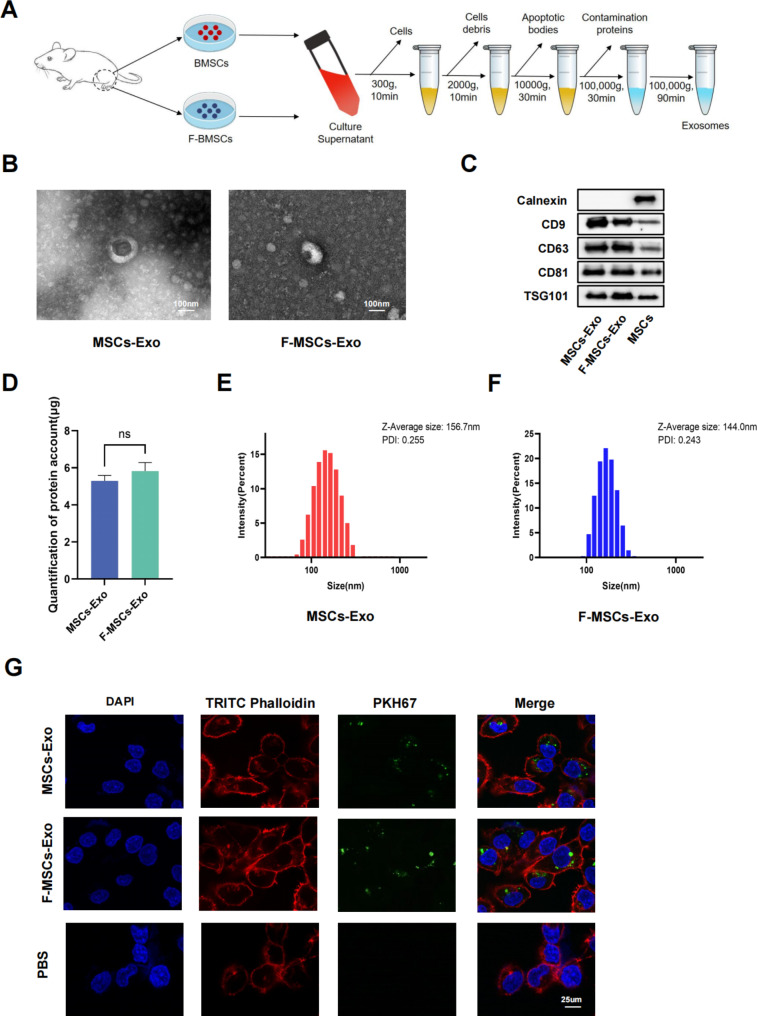
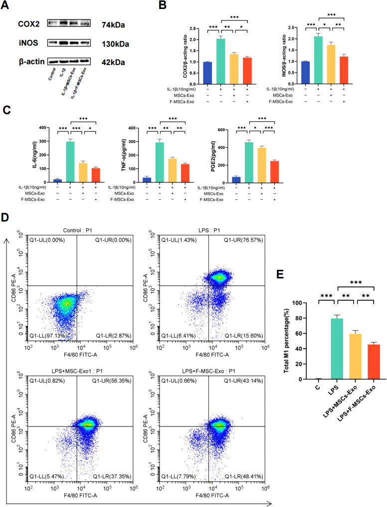
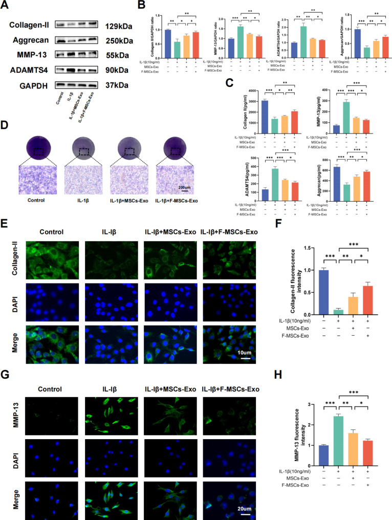
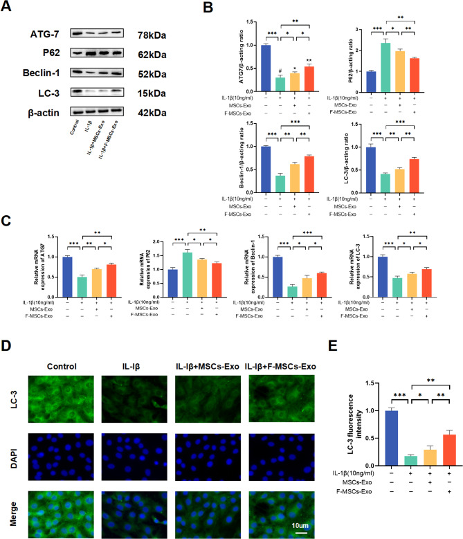
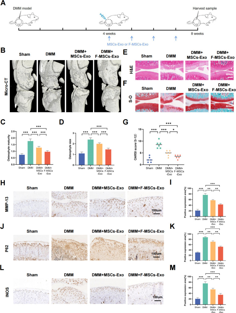
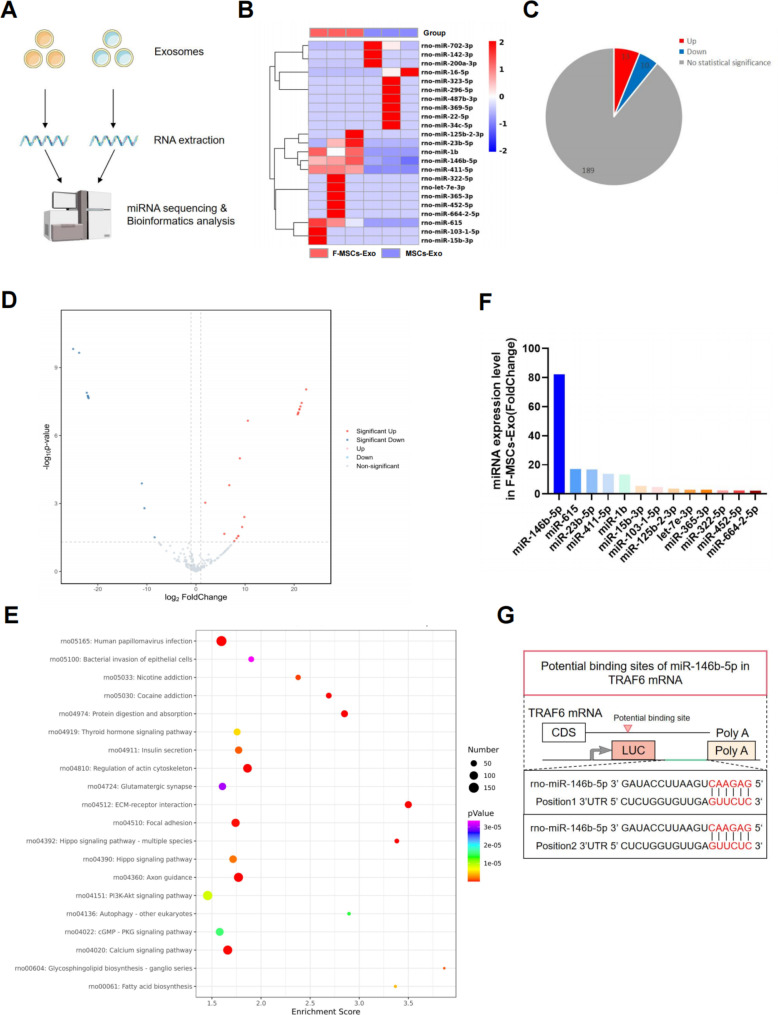
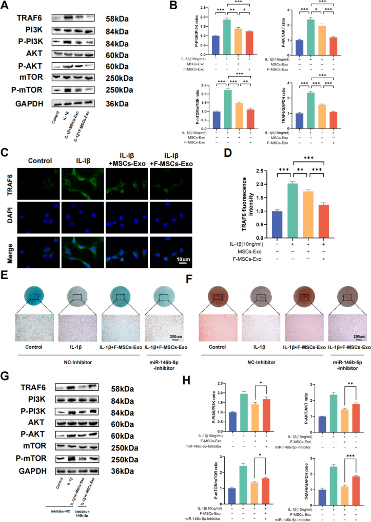
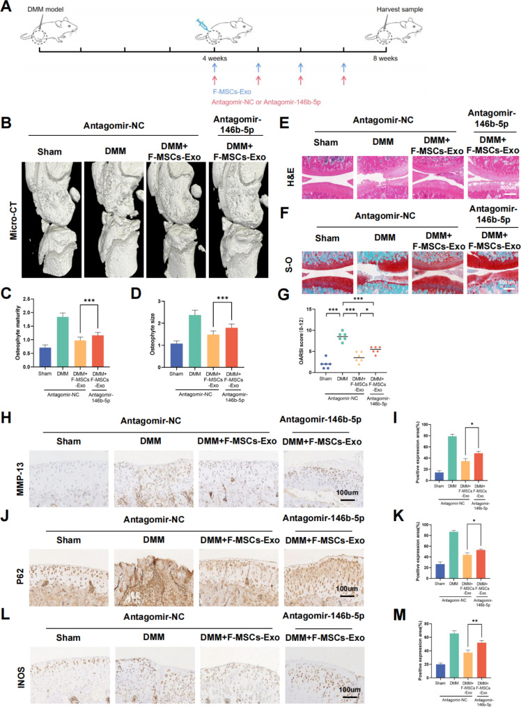
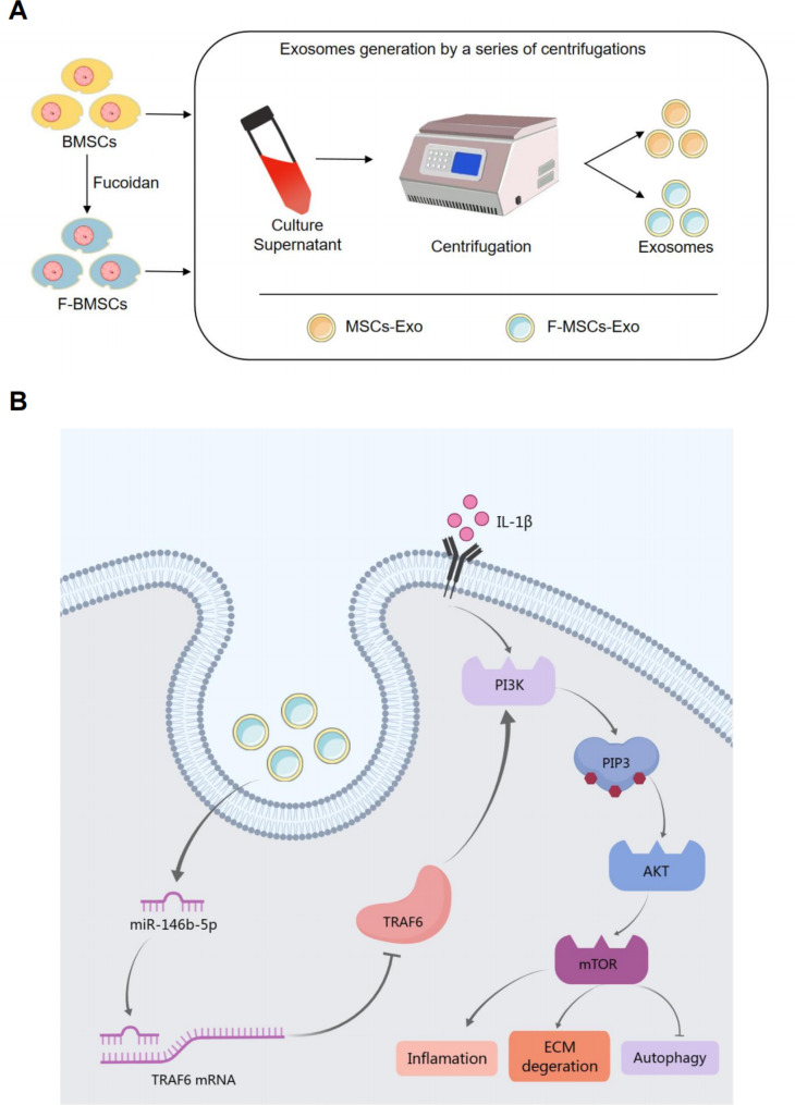
Osteoarthritis (OA) is a common degenerative joint disease characterized by progressive cartilage degradation and inflammation. In recent years, mesenchymal stem cells (MSCs) derived exosomes (MSCs-Exo) have attracted widespread attention for their potential role in modulating OA pathology. However, the unpredictable therapeutic effects of exosomes have been a significant barrier to their extensive clinical application. In this study, we investigated whether fucoidan-pretreated MSC-derived exosomes (F-MSCs-Exo) could better protect chondrocytes in osteoarthritic joints and elucidate its underlying mechanisms. In order to evaluate the role of F-MSCs-Exo in osteoarthritis, both in vitro and in vivo studies were conducted. MiRNA sequencing was employed to analyze MSCs-Exo and F-MSCs-Exo, enabling the identification of differentially expressed genes and the exploration of the underlying mechanisms behind the protective effects of F-MSCs-Exo in osteoarthritis. Compared to MSCs-Exo, F-MSCs-Exo demonstrated superior effectiveness in inhibiting inflammatory responses and extracellular matrix degradation in rat chondrocytes. Moreover, F-MSCs-Exo exhibited enhanced activation of autophagy in chondrocytes. MiRNA sequencing of both MSCs-Exo and F-MSCs-Exo revealed that miR-146b-5p emerged as a promising candidate mediator for the chondroprotective function of F-MSCs-Exo, with TRAF6 identified as its downstream target. In conclusion, our research results demonstrate that miR-146b-5p encapsulated in F-MSCs-Exo effectively inhibits TRAF6 activation, thereby suppressing inflammatory responses and extracellular matrix degradation, while promoting chondrocyte autophagy for the protection of osteoarthritic cartilage cells. Consequently, the development of a therapeutic approach combining fucoidan with MSC-derived exosomes provides a promising strategy for the clinical treatment of osteoarthritis.

Keywords: Exosomes; Fucoidan; MSCs; Osteoarthritis; TRAF6; miR-146b-5p.


Conflict of interest statement

The authors declare no competing interests.

Figures

Fig. 1
Fig. 1
Isolation and identification of MSCs-Exo and F-MSCs-Exo. (A) Schematic diagram of obtaining MSCs-Exo and F-MSCs-Exo by ultracentrifugation. (B) TEM analysis of the morphology of MSCs-Exo and F-MSCs-Exo. (C) Western blot evaluation of surface markers of MSCs-Exo and F-MSCs-Exo. (D) Quantification of protein concentration by BCA method. (E, F) The particle size and zeta potential of MSCs-Exo and F-MSCs-Exo were analyzed by DLS method. (G) Cellular internalization of MSCs-Exo and F-MSCs-Exo. (ns, no significant difference; *p < 0.05; **p < 0.01; ***p < 0.001; n = 3)
Fig. 2
Fig. 2
Effects of MSCs-Exo and F-MSCs-Exo on inflammatory response and M1 polarization in vitro. (A, B) Western blot analysis was performed to detect the effects of MSCs-Exo and F-MSCs-ExoOD on inflammatory factors in chondrocytes induced by IL-1β. (C) The levels of IL-6, TNF-α, and PGE2 in the chondrocyte culture supernatant after IL-1β induction were measured using ELISA kits to assess the impact of MSCs-Exo and F-MSCs-Exo on these inflammatory mediators. (D, E) Flow cytometry was used to investigate the influence of MSCs-Exo and F-MSCs-Exo on M1 polarization. (ns, no significant difference; *p < 0.05; **p < 0.01; ***p < 0.001; n = 3)
Fig. 3
Fig. 3
Effects of MSCs-Exo and F-MSCs-Exo on the synthesis and metabolism of cartilage extracellular matrix in vitro. (A, B) Western blot analysis was performed to detect the impact of MSCs-Exo and F-MSCs-Exo on cartilage extracellular matrix synthesis and metabolism markers. (C) ELISA kits were used to measure the levels of Collagen II, Aggrecan, MMP-13, and ADAMTS-4 in the cell culture supernatant. (D) Cartilage extracellular matrix was directly visualized using toluidine blue staining (scale bar = 200 μm). (E ,F) The expression of Collagen II was quantitatively analyzed using immunofluorescence staining and ImageJ software (scale bar = 10 μm). (G, H) The expression of MMP-13 was quantitatively analyzed using immunofluorescence staining and ImageJ software (scale bar = 20 μm). (ns, no significant difference; *p < 0.05; **p < 0.01; ***p < 0.001; n = 3)
Fig. 4
Fig. 4
Effects of MSCs-Exo and F-MSCs-Exo on autophagy of chondrocytes in vitro. (A, B) Western blot analysis was performed to detect the impact of MSCs-Exo and F-MSCs-Exo on autophagy-related indicators of chondrocytes. (C) Real-time PCR technology was used to evaluate the effects of MSCs-Exo and F-MSCs-Exo on autophagy-related indicators of chondrocytes at the gene expression level. (D, E) The expression of LC-3, a marker for autophagy, was quantitatively analyzed using immunofluorescence staining and ImageJ software to provide a detailed assessment of the autophagy levels in response to MSCs-Exo and F-MSCs-Exo treatment (scale bar = 10 μm). (ns, no significant difference; *p < 0.05; **p < 0.01; ***p < 0.001; n = 3)
Fig. 5
Fig. 5
Protective effects of MSCs-Exo and F-MSCs-Exo on osteoarthritis in rats. (A) A schematic diagram was provided, illustrating how MSCs-Exo and F-MSCs-Exo were used to treat osteoarthritis in rats. (B) Micro-CT 3D reconstruction of rat knee joints was conducted to visualize the structural changes and alterations in the knee join. (C, D) Quantitative analysis of the micro-CT results was performed to assess the changes in the knee joint’s structure. (E, F) H-E staining and S-O staining of rat knee joint sections at 8 weeks after operation were conducted for histological examination (scale bar = 500 μm). (G) The Osteoarthritis Research Society International (OARSI) score was used to evaluate the severity of osteoarthritis in rat cartilage, n = 6. (H to M) Immunohistochemical analysis was performed to visualize and quantify the expression levels of MMP-13, P62, and INOS in the rat knee joint (scale bar = 100 μm). (ns, no significant difference; *p < 0.05; **p < 0.01; ***p < 0.001; n = 3)
Fig. 6
Fig. 6
miR-146b-5p is a candidate effector for F-MSCs-Exo-mediated improvement in osteoarthritis. (A) A schematic diagram was presented to illustrate the process of miRNA sequencing and subsequent bioinformatics analysis. (B) The heat map showed the differential expression profile of miRNAs between MSCs-Exo and F-MSCs-Exo. (C, D) The pie chart and volcano plot displayed the distribution of up-regulated and down-regulated miRNAs between MSCs-Exo and F-MSCs-Exo. (E) KEGG enrichment analysis of F-MSCs-Exo was performed to investigate the potential biological pathways and processes affected by these miRNAs in the treatment of osteoarthritis. (F) Expression of up-regulated miRNAs in F-MSCs-Exo. (G) Targetscan was used to predict the binding site of rat miR-146b-5p and TRAF6
Fig. 7
Fig. 7
Enriched miR-146b-5p in F-MSCs-Exo inhibits PI3K/AKT/mTOR pathway by targeting TRAF6. (A, B) Western blot analysis was performed to detect the impact of F-MSCs-Exo on TRAF6 and the PI3K/AKT/mTOR pathway in rat chondrocytes. (C, D) The expression of TRAF6 was quantitatively analyzed using immunofluorescence staining and ImageJ software (scale bar = 10 μm). (E, F) Direct visualization of chondrocytes treated with nc-inhibitor and miR-146b-5p-inhibitor was performed using Alcian blue staining and safranin staining. (G, H) Western blot analysis was conducted to examine the expressions of TRAF6 and the PI3K/AKT/mTOR pathway in chondrocytes after treatment with nc-inhibitor and miR-146b-5p-inhibitor. (ns, no significant difference; *p < 0.05; **p < 0.01; ***p < 0.001; n = 3)
Fig. 8
Fig. 8
Antagomir-146b-5p reverses the therapeutic effect of F-MSCs-Exo on osteoarthritis in rats. (A) Schematic diagram of the experiment evaluating whether miR-146b-5p is involved in the treatment of osteoarthritis in rats by F-MSCs-Exo. (B) Micro-CT 3D reconstruction of the rat knee joint was performed to visualize structural changes and alterations caused by Antagomir-146b-5p treatment. (C, D) Quantitative analysis of micro-CT results. (E, F) H-E staining and S-O staining of rat knee joint sections were conducted for histological examination, enabling the assessment of tissue morphology and cartilage integrity (scale bar = 500 μm). (G) The Osteoarthritis Research Society International (OARSI) score was used to evaluate the severity of osteoarthritis in rat cartilage, n = 6. (H to M) Immunohistochemical analysis was performed to visualize and quantify the expression levels of MMP-13, P62, and INOS in the rat knee joint after Antagomir-146b-5p treatment (scale bar = 100 μm). (ns, no significant difference; *p < 0.05; **p < 0.01; ***p < 0.001; n = 3)
Fig. 9
Fig. 9
Schematic diagram of the protective effect of F-MSCs-Exo on OA. (A) Obtain MSCs-Exo and F-MSCs-Exo by ultracentrifugation. (B) Enriched miR-146b-5p in F-MSCs-Exo plays a role in the treatment of osteoarthritis by targeting TRAF6 and inhibiting PI3K/AKT/mTOR signaling pathway

References

    1. Katz JN, Arant KR, Loeser RF. Diagnosis and treatment of hip and knee osteoarthritis: a review. JAMA. 2021;325(6):568–78. doi: 10.1001/jama.2020.22171. - DOI - PMC - PubMed
    1. Goldring MB, Goldring SR, Osteoarthritis J Cell Physiol. 2007;213(3):626–34. doi: 10.1002/jcp.21258. - DOI - PubMed
    1. Quicke JG, Conaghan PG, Corp N, Peat G. Osteoarthritis year in review 2021: epidemiology & therapy. Osteoarthritis Cartilage. 2022;30(2):196–206. doi: 10.1016/j.joca.2021.10.003. - DOI - PubMed
    1. Block JA, Cherny D. Management of knee osteoarthritis: what internists need to know. Med Clin North Am. 2021;105(2):367–85. doi: 10.1016/j.mcna.2020.10.005. - DOI - PubMed
    1. Goudarzi R, Dehpour AR, Partoazar A. Nanomedicine and regenerative medicine approaches in osteoarthritis therapy. Aging Clin Exp Res. 2022;34(10):2305–15. doi: 10.1007/s40520-022-02199-5. - DOI - PubMed