

Histopathological Interpretation of TMJ Osteophyte: Report and Review
- PMID: 38105841
- PMCID: PMC10719170
- DOI: 10.1007/s12663-023-01938-z
Histopathological Interpretation of TMJ Osteophyte: Report and Review
Abstract
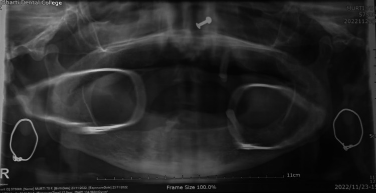
Introduction: Osteophytes are osseocartilaginous structures which are commonly found in common degenerative joint diseases. It can be free or attached to nature. There is a paucity of information in the literature regarding the histopathological interpretation of osteophytes in Temporomandibular Joint (TMJ).
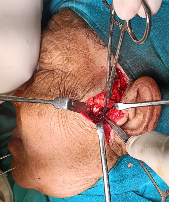
Purpose: To report the incidental finding of osteophytes in cases of chronic protracted TMJ dislocation.
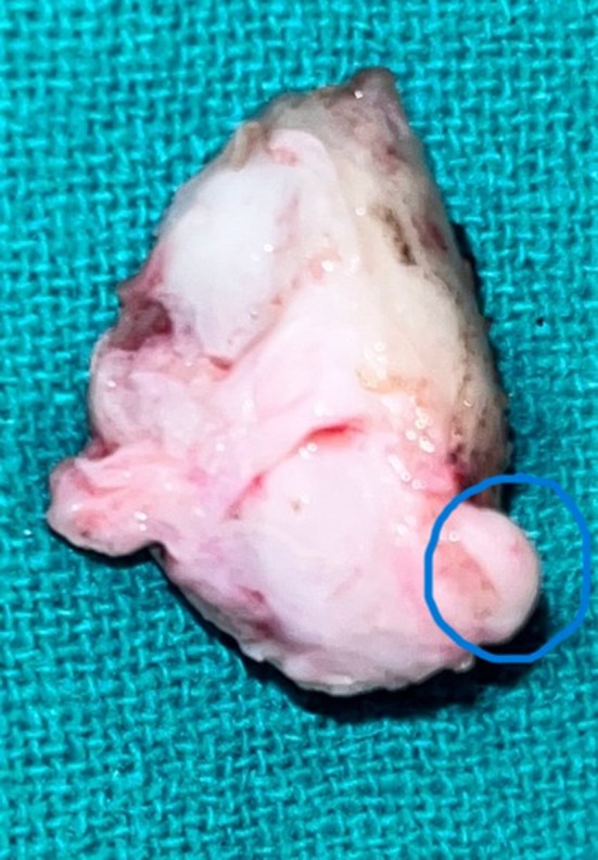
Material and method: To present case of an elderly 72-year-old female and a 35-year-old male with chronic protracted TMJ dislocation who were treated surgically for their condition with an incidental finding of an osteophyte in TMJ intraoperatively.
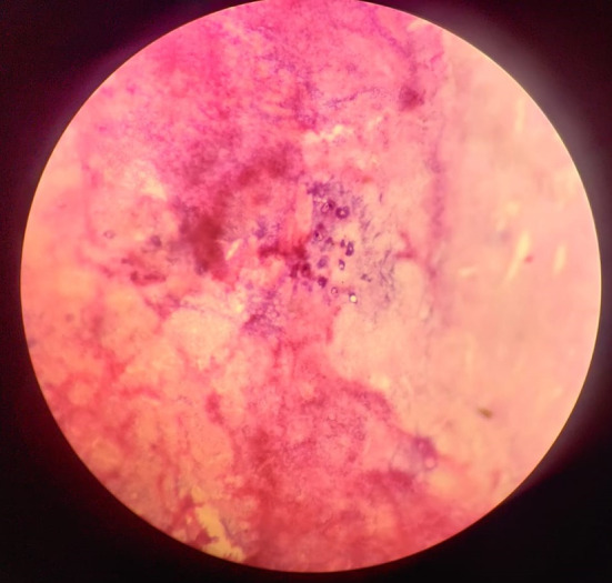
Result: The histopathological examination revealed fibrocartilaginous core tissue surrounded by bone formation due to dystrophic calcification.
Conclusion: The microscopic examination is mandatory in order to study and understand the pathophysiology of an osteophyte. This study presents rare histopathological evidence of an osteophyte. It reflects the process of its formation and the possibilities of fate of an osteophyte in TMJ.
© The Association of Oral and Maxillofacial Surgeons of India 2023.
Figures


References
-
- Petrikowski CG. Diagnostic imaging of temporomandibular joint. In: White SC, Pharoah MJ, editors. Oral radiology: principles and interpretation. 6. Jordan Hill, Oxford: Elsevier Mosby Inc; 2008. pp. 473–502.
Publication types
LinkOut - more resources
Full Text Sources