Review
. 2023 Dec;22(4):749-761.
doi: 10.1007/s12663-023-02053-9. Epub 2023 Dec 5.

Facial Asymmetry-Demystifying the Entity

Affiliations
Review

Facial Asymmetry-Demystifying the Entity

Nalla Viveka Vardhan Reddy et al. J Maxillofac Oral Surg. 2023 Dec.

Abstract

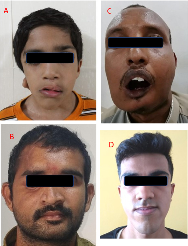
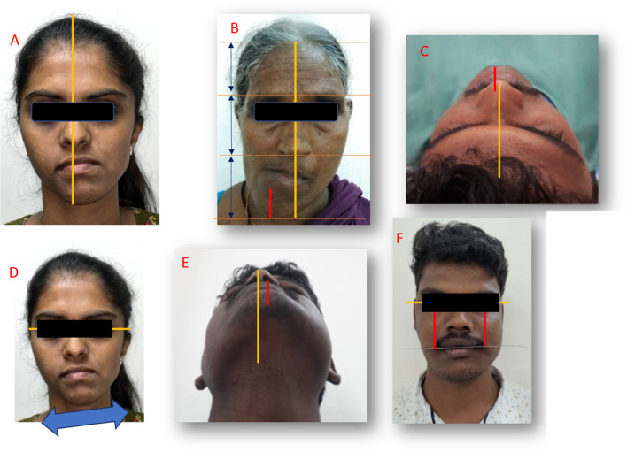
Introduction/background: Perfect facial symmetry has always been considered a hallmark of beauty, but, is almost elusive in nature. However, clinically evident skeletal facial asymmetry on the other hand is quite common, which can result from congenital deformities, developmental abnormalities, secondary to maxillofacial trauma and it is an entity maxillofacial surgeons deal with on a regular basis. Surgical correction of facial asymmetry is challenging, as it not only involves the correction of the skeletal asymmetry for an aesthetic outcome, but, also the improvement of the soft tissue drape and dental occlusal harmony. This results in rehabilitation of functional components of orofacial complex like speech, deglutition and phonation.

Objective: With this paper, we intend to throw a light on this challenging aspect of maxillofacial surgery, along with giving the next generation of maxillofacial surgeons a direction to explore the topic further.

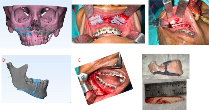
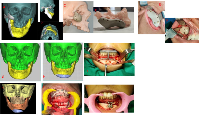
Conclusion: Meticulous evaluation and diagnosis of the patient's problems with latest diagnostic methods like 3-dimensional imaging and surgical treatment with orthognathic surgery, gap arthroplasty or distraction osteogenesis, utilizing cutting edge 3-D virtual planning will result in better outcomes.This review will collate the information available in the literature, along with the authors' recommendations for better planning and execution of this challenging puzzle of facial asymmetry.

Keywords: 3-D virtual planning; Facial asymmetry; Orthognathic surgery; Soft tissue augmentation.


Figures

Fig. 1
Fig. 1
A (congenital or prenatal asymmetry) B (acquired from an injury) C (acquired from disease) D (developmental)
Fig. 2
Fig. 2
A (Frontal view showing true facial midline for documenting facial asymmetry with deviation of mandible toward left side) B (Frontal view showing the difference in facial heights based on thirds and increased lower third contributing to the asymmetry) C (Bird’s eye view demonstrating asymmetry) D (frontal view illustrating cant of lower border of mandible) E (submental view documenting mandibular asymmetry) F (frontal view showing cant of occlusal plane to interpupillary plane)
Fig. 3
Fig. 3
AC (Differential impaction of maxilla to correct the cant planned virtually and 3D cutting guides used to simulate the virtual plan) DE (subtraction of over-projected side, the angle and lower border for asymmetry correction with the aid of cutting guide)
Fig. 4
Fig. 4
AD (Correction of the under-projecte 4D side of the asymmetry with a customized implant) E, F (intra-operative images of the implant positioned as per the virtual plan) G, H (virtual planning to correct the chin asymmetry) I (cutting guide to accurately perform the osteotomy of chin and position it as per the plan) J (Pre-bent plate used to accurately advance the chin to the pre-planned desired position) K, L (Cant correction of the chin by differential down-fracture and bone grafting the osteotomy gap)
Fig. 5
Fig. 5
A, B (a case of right Hemifacial Microsomia-a congenital form of facial asymmetry involving skeletal, dental, and soft tissue components) C (OPG demonstrating the hypoplastic right condyle) D, E (3D CT reconstruction and STL model) F, G (post distraction osteogenesis) H (post distraction OPG) I, J (Bony augmentation with custom PEEK implant to correct the soft tissue deformity). K, L (post distractor removal, genioplasty, and soft tissue augmentation)
Fig. 6
Fig. 6
A, B (pre-operative clinical image and OPG of right hemimandibular elongation) C, D (post-operative clinical image and OPG) E (maxillary osteotomy for differential impaction) F (intraoral condylar shaving) G (resected condylar segment)
Fig. 7
Fig. 7
A, B (pre and post-op frontal images of right condylar hyperplasia) C (3D reconstruction showing right condylar hyperplasia) D (post-op OPG) EH (pre and post-operative frontal and worm's view images of right mandibular hyperplasia) I, J (pre and post-op OPG)
Fig. 8
Fig. 8
A (pre-op frontal image of left condylar osteochondroma leading to facial asymmetry) B (severely deranged dental occlusion with mandible midline shifted to the right) C, D (pre-op imaging) E (post-op frontal image) F (post-op and post orthodontics dental occlusion after debonding) G (post-op OPG) H, I (intra-op images of right condylectomy done extra orally) J (right sagittal split osteotomy done to correct the deviated mandibula midline and the occlusion)
Fig. 9
Fig. 9
A, B, (pre-op frontal and profile images of facial asymmetry resulting from hemi mandibular left ramus shortening post ankylotic release) C, D (post-op frontal and profile images of asymmetry correction with unilateral simultaneous maxillo- mandibular DO) E, F (pre-op and post-op OPG) G, H (pre-op and post-op lateral cephalogram demonstrating improvement in profile and upper airway) I, J (pre-op and post-op frontal images of facial asymmetry due to left ramus shortening post ankylotic release corrected with unilateral mandibular DO) K (left mandibular distractor in position)
Fig. 10
Fig. 10
A, B (pre-op frontal and profile view of residual deformity at left lower border of mandible following orthognathic surgery of mandibular setback, maxillary advancement, and genioplasty in a patient with asymmetric prognathic mandible) CE (virtual planning illustrating augmentation of the lower border with patient-specific PEEK implant) F, G (post-op images) I (post-op OPG)

References

    1. Lindauer SJ. Asymmetries: diagnosis and treatment (editorial) Semin Orthod. 1998;4(3):133. doi: 10.1016/S1073-8746(98)80013-3. - DOI - PubMed
    1. Thiesen G, Gribel BF, Freitas MPM. Facial asymmetry: a current review. Dental Press J Orthod. 2015;20(6):110–125. doi: 10.1590/2177-6709.20.6.110-125.sar. - DOI - PMC - PubMed
    1. Cheong YW, Lo LJ. Facial asymmetry: etiology, evaluation, and management. Chang Gung Med J. 2011;34(4):341–351. - PubMed
    1. Nanda R, Margolis MJ. Treatment strategies for midline discrepancies. Semin Orthod. 1996;2(2):84–89. doi: 10.1016/S1073-8746(96)80046-6. - DOI - PubMed
    1. Burstone CJ. Diagnosis and treatment planning of patients with asymmetries. Semin Orthod. 1998;4(3):153–164. doi: 10.1016/S1073-8746(98)80017-0. - DOI - PubMed

LinkOut - more resources