Bulldog Scalp Syndrome
- PMID: 38105879
- PMCID: PMC10721373
- DOI: 10.1055/s-0043-1776897
Bulldog Scalp Syndrome
Abstract
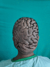
Bulldog scalp syndrome or cutis verticis gyrata (CVG) is a rare cutaneous disorder with an incidence of just 0.026 to 1 per 100,000 population and cosmetic problems should not be ignored as they can affect the quality of life of patients in social and psychological aspects. In CVG the scalp thickens to form folds resembling sulci and gyri just as the skin fold of bulldog. It is a clinical diagnosis with various etiologies. It is classified as primary essential or nonessential and secondary CVG. It can manifest with symptoms ranging from mild to severe intensity. Cosmetic problems are the major concern that can affect patients' social and psychological health. If the folds are heavy, they can cause mass symptoms. Thus, surgery remains the definitive treatment option for improving the cosmetic appearance. Both our cases have different etiologies, however, were managed surgically with removal of skin folds (gyrae) and scoring of aponeuroses of the scalp followed by stretching of the scalp and closure to improve appearance. The surgical team as well as patients were satisfied with the appearance of the scalp after healing. CVG though a rare disease with various etiologies is a benign condition with good prognosis with no reports of malignant transformation so far.
Keywords: bulldog scalp; cutis verticis gyrata; scalp flap.
Association of Plastic Surgeons of India. This is an open access article published by Thieme under the terms of the Creative Commons Attribution-NonDerivative-NonCommercial License, permitting copying and reproduction so long as the original work is given appropriate credit. Contents may not be used for commercial purposes, or adapted, remixed, transformed or built upon. ( https://creativecommons.org/licenses/by-nc-nd/4.0/ ).
Conflict of interest statement
Conflict of Interest None declared.
Figures


References
-
- Shareef S, Horowitz D, Kaliyadan F.Cutis Verticis Gyrata 2023. Jul 10. In: StatPearls [Internet].Treasure Island (FL)StatPearls Publishing; 2023 Jan - PubMed
-
- Larsen F, Birchall N. Cutis verticis gyrata: three cases with different aetiologies that demonstrate the classification system. Australas J Dermatol. 2007;48(02):91–94. - PubMed
-
- Schöttler L, Denisjuk N, Körber A, Grabbe S, Dissemond J. Asymptomatic cerebriform folds of the scalp. Diagnosis: primary cutis verticis gyrata (CVG) [in German] Hautarzt. 2007;58(12):1058–1061. - PubMed
Publication types
LinkOut - more resources
Full Text Sources

