Review
. 2023 Nov 30:17:1277399.
doi: 10.3389/fnins.2023.1277399. eCollection 2023.

Immunology of amyotrophic lateral sclerosis - role of the innate and adaptive immunity

Affiliations
Review

Immunology of amyotrophic lateral sclerosis - role of the innate and adaptive immunity

Stefan Mimic et al. Front Neurosci. .

Abstract

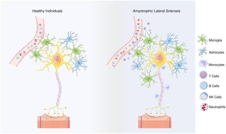
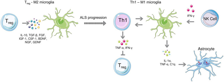
This review aims to summarize the latest evidence about the role of innate and adaptive immunity in Amyotrophic Lateral Sclerosis (ALS). ALS is a devastating neurodegenerative disease affecting upper and lower motor neurons, which involves essential cells of the immune system that play a basic role in innate or adaptive immunity, that can be neurotoxic or neuroprotective for neurons. However, distinguishing between the sole neurotoxic or neuroprotective function of certain cells such as astrocytes can be challenging due to intricate nature of these cells, the complexity of the microenvironment and the contextual factors. In this review, in regard to innate immunity we focus on the involvement of monocytes/macrophages, microglia, the complement, NK cells, neutrophils, mast cells, and astrocytes, while regarding adaptive immunity, in addition to humoral immunity the most important features and roles of T and B cells are highlighted, specifically different subsets of CD4+ as well as CD8+ T cells. The role of autoantibodies and cytokines is also discussed in distinct sections of this review.

Keywords: adaptive immune system; amyotrophic lateral sclerosis; innate immune system; neurodegeneration; neuroimmunology and neuropathy.


Conflict of interest statement

The authors declare that the research was conducted in the absence of any commercial or financial relationships that could be construed as a potential conflict of interest.

Figures

Figure 1
Figure 1
Differences between healthy individuals and ALS patients regarding the types of inflammatory cells. Members of the adaptive innate immune system, T and B cells play role in ALS progression, though their involvement may depend on the stage of the disease. Microglia and astrocytes are considered as the main contributors to the non-cell autonomous mechanism in ALS. The neuroprotective M2 microglia provide protection in the beginning of the disease by releasing anti-inflammatory mediators. However, as the disease progresses, a transition from the M2 to the neurotoxic M1 phenotype is observed. Microglial polarization in ALS is regulated with T cells and astrocytes’ responses. In terms of the first, anti-inflammatory cytokines IL-4 and IL-10 are released by Th2 cells and Tregs, during the early stage of disease, in addition, Th2 cells also secrete various neurotrophic factors while as the disease progresses, Th1 cells release the pro-inflammatory mediators and contribute to the M1 polarization. Similarly, the NF-κB activation in spinal cord astrocytes can promote the neuroprotective phenotype of microglia and inhibit disease progression, but prolonged NF-κB activation in the later stages of the disease promotes pro-inflammatory microglial responses. In addition, impairments in glutamate transporters limits glutamate uptake of astrocytes, ultimately leading to dysfunction of motoneurons. Besides microglia and astrocytes, NK cells and monocytes can infiltrate the region and contribute to the inflammatory response. In the peripheral blood of the ALS patients, neutrophil percentages were reported to be increased, and the increase in their ratio to lymphocytes correlates with the disease progression. By releasing hematopoietic serine proteases, neutrophils also regulate NK cell toxicity.
Figure 2
Figure 2
Microglial polarization during early (left) and late stages (right) of ALS. Anti-inflammatory microglia protect motor neurons at the beginning of the disease, while a transition to the neurotoxic M1 phenotype is observed as the disease progresses. T cell-microglia interaction plays role in microglial polarization, NK cells also contribute to the M1 polarization by secreting IFN-γ. Activated microglia release, IL-1α, TNF-α as and C1q which induce neurotoxic responses of astrocytes.
Figure 3
Figure 3
Even though ALS is considered as motor neuron disease, not only neurons but glial cells including astrocytes also play a role in the non-cell autonomous disease pathology (Stoklund Dittlau and Van Den Bosch, 2023). Under physiological conditions, astrocytes are involved in the modulation of neuronal synapse plasticity; ion and metabolic homeostasis; cellular communication; supporting blood–brain barrier and providing structural support. In healthy tissues, non-activated astrocytes remain immobile and support neuronal homeostasis while upon damage, they activate and initiate an immune response to fight against the harmful agents together with microglia. In neurodegenerative diseases including ALS, an abnormal astrocyte reactivity that contributes to neuronal death is observed. Being the most abundant excitatory neurotransmitter in the nervous system, glutamate is found at high concentrations in pre-synaptic nerve terminals where it binds to receptors such as α-amino-3-hydroxy-5-methy-4-isoxazolepropionic acid (AMPA) and N-methyl-D-aspartate (NMDA) receptors. Since high levels of glutamate may lead to a lethal increase in intracellular calcium astrocytes play a protective role in glutamate clearance from the synaptic cleft via excitatory amino acid transporters (EAATs). Among five EAATs identified, EAAT-1 and EAAT-2 are primarily expressed on astrocytes (Ng and Ng, 2022). Dysregulation of the human counterpart of GLT1, EAAT-2 (Rimmele and Rosenberg, 2016) expression has been shown to be altered in ALS and reported to contribute increased glutamate levels in the cerebrospinal fluid of ALS patients. Moreover, astrocytes produce inflammatory mediators that contribute to neuronal death (Stoklund Dittlau and Van Den Bosch, 2023). Mitochondrial dysfunction in astrocytes also promote neuronal death in ALS (Zhao et al., 2022).

References

    1. Abel A. M., Yang C., Thakar M. S., Malarkannan S. (2018). Natural killer cells: development, maturation, and clinical utilization. Front. Immunol. 9:1869. doi: 10.3389/fimmu.2018.01869, PMID: - DOI - PMC - PubMed
    1. Abonia J. P., Friend D. S., Austen W. G., Jr., Moore F. D., Jr., Carroll M. C., Chan R., et al. . (2005). Mast cell protease 5 mediates ischemia-reperfusion injury of mouse skeletal muscle. J. Immunol. 174, 7285–7291. doi: 10.4049/jimmunol.174.11.7285, PMID: - DOI - PMC - PubMed
    1. Allen N. J., Lyons D. A. (2018). Glia as architects of central nervous system formation and function. Science 362, 181–185. doi: 10.1126/science.aat0473, PMID: - DOI - PMC - PubMed
    1. Andjus P. R., Bataveljić D., Vanhoutte G., Mitrecic D., Pizzolante F., Djogo N., et al. . (2009). In vivo morphological changes in animal models of amyotrophic lateral sclerosis and Alzheimer's-like disease: MRI approach. Anat. Rec. (Hoboken) 292, 1882–1892. doi: 10.1002/ar.20995 - DOI - PubMed
    1. Andjus P. R., Khiroug L., Nistri A., Cherubini E. (1996). ALS IgGs suppress [Ca2+]i rise through P/Q-type calcium channels in central neurones in culture. Neuroreport 7, 1914–1916. doi: 10.1097/00001756-199608120-00008, PMID: - DOI - PubMed