[Preprint]. 2023 Dec 4:2023.12.03.569791.
doi: 10.1101/2023.12.03.569791.

Tumor-selective effects of active RAS inhibition in pancreatic ductal adenocarcinoma

Affiliations

Tumor-selective effects of active RAS inhibition in pancreatic ductal adenocarcinoma

Urszula N Wasko et al. bioRxiv. .

Abstract

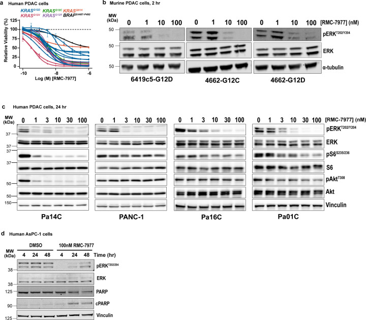
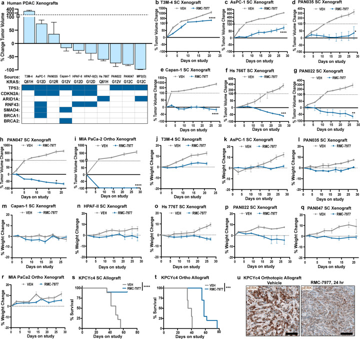
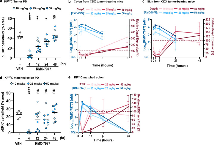
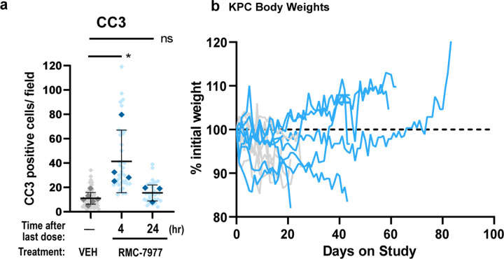
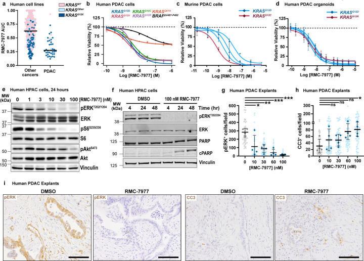
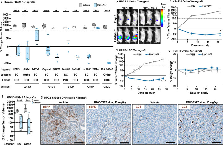
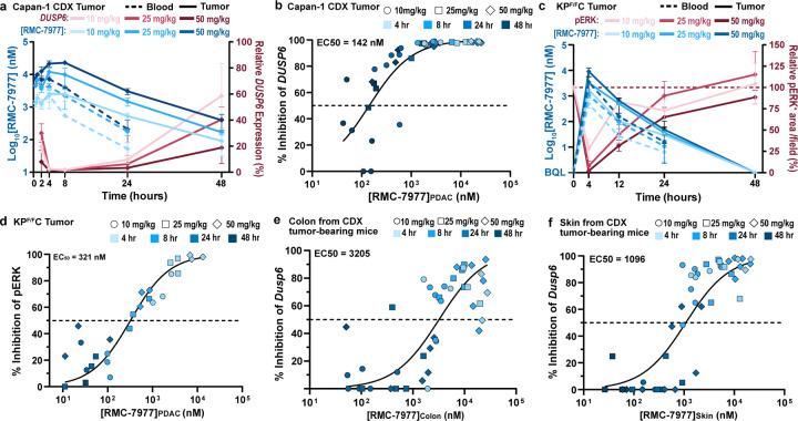
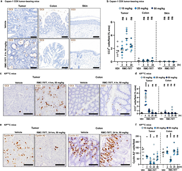
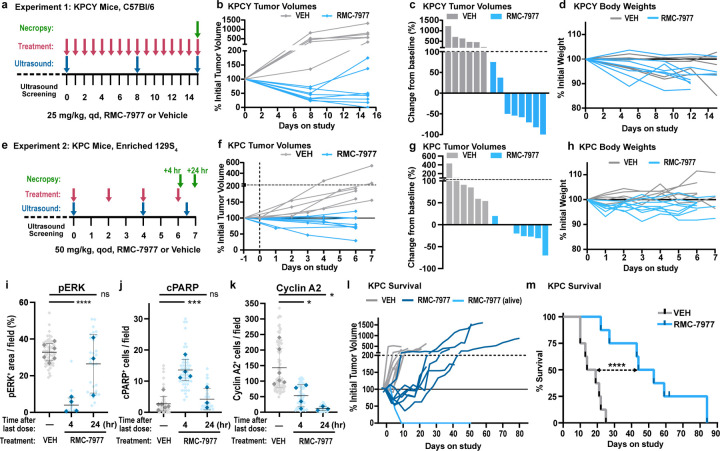
Broad-spectrum RAS inhibition holds the potential to benefit roughly a quarter of human cancer patients whose tumors are driven by RAS mutations. However, the impact of inhibiting RAS functions in normal tissues is not known. RMC-7977 is a highly selective inhibitor of the active (GTP-bound) forms of KRAS, HRAS, and NRAS, with affinity for both mutant and wild type (WT) variants. As >90% of human pancreatic ductal adenocarcinoma (PDAC) cases are driven by activating mutations in KRAS, we assessed the therapeutic potential of RMC-7977 in a comprehensive range of PDAC models, including human and murine cell lines, human patient-derived organoids, human PDAC explants, subcutaneous and orthotopic cell-line or patient derived xenografts, syngeneic allografts, and genetically engineered mouse models. We observed broad and pronounced anti-tumor activity across these models following direct RAS inhibition at doses and concentrations that were well-tolerated in vivo. Pharmacological analyses revealed divergent responses to RMC-7977 in tumor versus normal tissues. Treated tumors exhibited waves of apoptosis along with sustained proliferative arrest whereas normal tissues underwent only transient decreases in proliferation, with no evidence of apoptosis. Together, these data establish a strong preclinical rationale for the use of broad-spectrum RAS inhibition in the setting of PDAC.


Conflict of interest statement

Competing interests J.J., Y.W., B.L., M.M., S.C., L.J., X.W., Y.C.Y., C.H., H.C., Y.G., R.Z., E.Q., Z.W., J.A.M.S., M.H., D.W., and M.S. are employees and stockholders of Revolution Medicines. R.H.V., B.Z.S., A.J.A., C.J.D., and K.P.O. received research funding from Revolution Medicines. A.J.A. consults for Anji Pharmaceuticals, Affini-T Therapeutics, Arrakis Therapeutics, AstraZeneca, Boehringer Ingelheim, Oncorus, Inc., Merck & Co. Inc., Mirati Therapeutics, Nimbus Therapeutics, Plexium, Revolution Medicines, Reactive Biosciences, Riva Therapeutics, Servier Pharmaceuticals, Syros Pharmaceuticals, T-knife Therapeutics, Third Rock Ventures, and Ventus Therapeutics. A.J.A. holds equity in Riva Therapeutics. A.J.A. has research funding from Bristol Myers Squibb, Deerfield, Inc., Eli Lilly, Mirati Therapeutics, Novartis, Novo Ventures, and Syros Pharmaceuticals. C.J.D. is a consultant/advisory board member for Cullgen, Deciphera Pharmaceuticals, Eli Lilly, Mirati Therapeutics, Reactive Biosciences, Revolution Medicines, Ribometrics, Sanofi, and SHY Therapeutics. C.J.D. has received research funding support from Deciphera Pharmaceuticals, Mirati Therapeutics, Reactive Biosciences, and SpringWorks Therapeutics. R.H.V. has received consulting fees from BMS, is an inventor on patents relating to cancer cellular immunotherapy, cancer vaccines, and KRAS immune epitopes, and receives royalties from Children’s Hospital Boston for a licensed research-only monoclonal antibody.

Figures

Extended Data Figure 1
(a) Viability levels of human PDAC cell lines with KRASG12X (AsPC-1, HPAC, Panc 05.04, PANC-1, Panc 10.05, PL45, SU.86.86, SW-1990, KP-4, HPAF-II, Capan-1, Capan-2, CFPAC-1, Panc 03.27, HuP-T4, MIA PaCa-2 and PSN-1), KRASQ61H (Hs 766T), and BRAFΔV487-P492 (BxPC-3) mutations treated with indicated concentrations of RMC-7977 for 4 hours. Data points represent the mean of technical replicates normalized to DMSO control. Error bars indicate s.d. KRAS mutations are indicated by curve colors. (b) Representative Western Blot images for three murine PDAC cell lines treated with DMSO or range of RMC-7977 concentrations (1–100 nM) for 2 hours. Protein levels of phospho-ERKT202/204 and total ERK were analyzed. Alpha-(α)-tubulin was used as loading control. (c) Representative Western Blot images for human PDAC cell lines treated with DMSO or range of RMC-7977 concentrations (1–100 nM) for 24 hours. Protein levels of phospho-ERKT202/204, total ERK, phospho-pS6S235/236 total S6, phospho-AktS473 and total Akt were analyzed. Vinculin was used as loading control. (d) Representative Western Blot images for AsPC-1 cell line treated with DMSO or RMC-7977 (100 nM) for indicated timepoints. Protein levels of phospho-ERKT202/204, total ERK, total PARP and cleaved PARP were analyzed. Vinculin was used as loading control.
Extended Data Figure 2
(a-k) Human PDAC xenograft models from Fig. 2. (a) Waterfall plot showing tumor volume change from baseline in RMC-7977 treated tumors. Error bars indicate ± s.e.m. Table shows selected genotypes for the xenograft panel with the row above indicating the KRAS mutation. Present co-mutations in each model shown as blue squares in the table. (b-i) Tumor growth curves for indicated xenograft models from (a), shown as percent tumor volume change from baseline over the course of treatment. Vehicle and RMC-7977 groups were compared by 2-way repeated measures ANOVA on the last measurement day of the vehicle group (*, p<0.05; **, p<0.01; ***, p<0.001; ****, p<0.0001). Error bars indicate ± s.e.m. (j-r) Tolerability of RMC-7977 as assessed by percent animal body weight change from baseline over the course of treatment, for indicated xenograft models from (a). Error bars indicate ± s.e.m. (s,t) Kaplan-Meier survival analysis comparing RMC-7977 and Vehicle treatment arms in (s) subcutaneous and (t) orthotopic KPCYc4 allograft models (***, p<0.001; ****, p<0.0001). (u) Representative IHC images of Vehicle and RMC-7977-treated KPCYc4 tumors collected at 24 hours post last dose and stained for phospho-ERKT202/204. Scale bars = 100 μm.
Extended Data Figure 3
(a,d) IHC analysis was ran on tissues collected from KPF/FC mice in Fig 3. (a) Quantification of phospho-ERKT202/204 IHC staining of tumors isolated from Vehicle and RMC-7977 treated KPF/FC mice. (b,c) PK/PD relationship in the colons and skin isolated from CDX tumor-bearing mice. Pharmacokinetic response shown as RMC-7977 concentration in (b) colon and (c) skin (solid blue lines) over time. Pharmacodynamic response shown as relative change in Dusp6 expression in colon or skin (red solid lines) over time. Shades of blue or red represent three tested doses. Error bars indicate s.d. (d) Quantification of phospho-ERKT202/204 IHC staining of colons isolated from Vehicle and RMC-7977-treated KPF/FC mice. (e) PK/PD relationship of RMC-7977 in the colons isolated from KPF/FC mice. Pharmacokinetic response shown as RMC-7977 concentration in colon (solid blue lines) over time. Pharmacodynamic response shown as relative change in pERK positive IHC staining in colon (red solid lines) over time. Shades of blue or red represent three tested doses. Error bars indicate s.d. (a,d) Analysis of IHC images based on 10–15 fields of view and plotted as average per tissue section. Shades of blue represent three tested doses. Results were compared by Student’s unpaired t-test (*, p<0.05; **, p<0.01; ****, p<0.0001).
Extended Data Figure 4
(a) IHC analysis of KPC tumors from Fig. 4. Tumors stained for CC3. Quantification of IHC images was compared between Vehicle, RMC-7977 (1 week + 4 hour) and RMC-7977 (1 week + 24 hour). Quantification was based on 10–15 fields of view (light shade), averaged per tumor (dark shade) and means were compared by Student’s unpaired t-test (*, p<0.05). Error bars indicate s.d. (b) Tolerability of RMC-7977 in KPC mice as assessed by percent animal body weight change from baseline over the course of treatment.
Fig 1.
Fig 1.. RMC-7977 demonstrates potent anti-tumor activity in in vitro and ex vivo models of PDAC.
(a) PRISM multiplex cell line screening testing changes in viability of 796 cancer cell lines in response to RMC-7977 treatment. Cell line viability was plotted as Area Under Curve (AUC) values. KRAS status indicated by color of the symbol. Horizontal lines indicate median. (b) Viability levels of human PDAC cell lines with KRASG12X (HPAF-II, HuP-T4, MIA PaCa-2 and PSN-1), KRASQ61H (Hs 766T), and BRAFΔV487-P492 (BxPC-3) mutations treated with indicated concentrations of RMC-7977 for 4 hours. (c) Viability levels of murine PDAC lines with KRASG12X (4662-G12D, 4662-G12C, 6419c5, 2838c3) mutations treated with indicated concentrations of RMC-7977 for 72 hours. (d) Viability levels of human PDAC organoids with KRASG12X mutations treated with indicated concentrations of RMC-7977 for 6 days. Data points in (b, c, d) represent the mean of technical 2–3 replicates normalized to DMSO control. Error bars indicate s.d. KRAS mutations are indicated by curve colors. (e) Western blots of HPAC cells treated with DMSO or range of RMC-7977 concentrations (1–100 nM) for 24 hours. Protein levels phospho-ERKT202/204, total ERK, phospho-pS6S235/236, total S6, phospho-AktS473 and total Akt were analyzed. Vinculin was used as loading control. (f) Western blots of HPAC cells treated with DMSO or RMC-7977 (100 nM) for indicated time points. Protein levels phospho-ERKT202/204, total ERK, total PARP and cleaved PARP were analyzed. Vinculin was used as loading control. (g-i) Ex vivo human PDAC explants treated with DMSO or a range of RMC-7977 concentrations (10 – 100 nM) for 24 hr (n=4). (g,h) Quantification of IHC images of explants stained for (g) phospho-ERKT202/204 and (h) CC3. Analysis based on 5–15 fields of view (light shade), averaged per explant slice (dark shade) compared by one-way ANOVA test with Tukey correction (*, p<0.05; **, p<0.01; ***, p<0.001). Error bars indicate s.d. (i) Representative IHC images of phosho-ERKT202/204 and CC3 staining of explants treated with DMSO or RMC-7977 (100 nM). Scale bars = 50 μm.
Fig 2.
Fig 2.. RMC-7977 exhibits anti-tumor activity in xenograft and allograft models of PDAC.
(a-e) Human PDAC xenograft models implanted either subcutaneously (SC) or orthotopically (Ortho) into immunodeficient mice. Tumor-bearing mice were treated with Vehicle or RMC-7977 (10 mg/kg, po, q.d.; n=3–10) for 21–28 days. (a) Boxplot showing percent change in tumor volumes at endpoint compared with baseline at Day 0 in Vehicle and RMC-7977 treatment arms. Each symbol represents one mouse. Source and format of each cell line, KRAS mutation, and tumor locations are indicated in the graph. Study arms were compared by Student’s unpaired t-test (*, p<0.05; **, p<0.01; ****, p<0.0001). (b) Representative bioluminescence images showing signal in HPAF-II orthotropic xenograft tumors at Day 0 and at Day 21 for Vehicle and RMC-7977 treatment arms. (c,d) Representative tumor growth curves for HPAF-II (c) orthotopic and (d) subcutaneous xenograft models treated with Vehicle or RMC-7977 (n=8/group), shown as percent tumor volume change from baseline over the course of treatment. Vehicle and RMC-7977 groups were compared by 2-way repeated measures ANOVA on the last measurement day of the vehicle group (*, p<0.05; ****p<0.0001). Error bars indicate ± s.e.m. (e) Tolerability of RMC-7977 as assessed by percent animal body weight change from baseline over the course of treatment. Error bars indicate ± s.e.m. (f, g) KPCY-derived PDAC cell line (6499c4) was transplanted either subcutaneously or orthotopically into syngeneic mice. Tumor-bearing mice were treated with Vehicle or RMC-7977 (10 mg/kg, po, q.d.; n=9–10/group). (f) Boxplot showing changes in subcutaneous and orthotopic tumor volumes at Day 14, compared with baseline at Day 0, in Vehicle and RMC-7977 treatment arms. Groups compared by Student’s unpaired t-test (***, p<0.001; ****, p<0.0001). Tumor locations as indicated in the graph. (g) Representative IHC images of Vehicle and RMC-7977-treated KPCY allograft tumors stained for phospho-ERKT202/204 and CC3. Scale bars = 100 μm.
Fig 3.
Fig 3.. Pharmacology of RAS addiction
(a,b) Capan-1 subcutaneous xenograft tumors were treated with Vehicle or RMC-7977 at 10 mg/kg, 25 mg/kg and 50 mg/kg. Tissues were harvested at indicated timepoints (n=3–6/timepoint/dose). (a) PK/PD in the Capan-1 xenograft model. Pharmacokinetic profile shown as RMC-7977 concentration in tumors (solid blue lines) and blood (dashed blue lines) over time. Pharmacodynamic response shown as relative change in DUSP6 mRNA expression (solid red lines). Shades of blue or red represent three tested doses. Values plotted as mean ± s.d. (b) PKPD relationship between RMC-7977 concentration and inhibition of DUSP6 expression in tumors. (c,d) Tumor-bearing KPF/FC mice were treated with RMC-7977 at 10 mg/kg, 25 mg/kg and 50 mg/kg. Tissues were harvested at indicated timepoints (n=3/timepoint/dose). (c) PK/PD in the tumors isolated from KPF/FC mice. Pharmacokinetic response shown as RMC-7977 concentration in tumors (solid blue lines) and blood (dashed blue lines) over time. Pharmacodynamic response shown as relative change in pERK positive IHC staining in tumors (solid red lines) over time. Shades of blue or red represent three tested doses. Values plotted as mean ± s.d. (d) PKPD relationship between RMC-7977 concentration and pERK inhibition in tumors. (e,f) PKPD relationship between RMC-7977 concentration and inhibition of Dusp6 expression in normal (e) colons and (f) skin, isolated from CDX tumor-bearing mice. (b,d,e,f) A 3-parameter sigmoidal exposure response model was fitted to the data to derive EC50 values. Timepoints represented by shades of blue and doses represented by symbol shapes.
Fig 4.
Fig 4.. Inhibition of RAS induces pancreatic tumor-selective apoptosis
(a) Representative IHC images of tumors, colons, and skin from Capan-1 xenograft model collected at 8 hours post single dose of Vehicle or RMC-7977, stained for CC3. Scale bars = 100 μm. (b) Quantification of CC3 staining in tumors, colon, and skin. (c) Representative IHC images of KPF/FC tumors and colons collected at 4 hours post single dose of Vehicle or RMC-7977, stained for CC3. Scale bars = 50 μm. (d) Quantification of CC3 staining in tumors and colons. (e) Representative IHC images of KPF/FC tumors and colons collected at 24 hours post single dose of Vehicle or RMC-7977, stained for Cyclin A2. Scale bars = 50 μm. (f) Quantification of Cyclin A2 staining in tumors and colons. (b,d,f) Analysis of IHC images based on 10–15 fields of view and plotted as average per tissue section. Shades of blue represent three tested doses. Results were compared by Student’s unpaired t-test (*, p<0.05; **, p<0.01; ***, p<0.001; ****, p<0.0001).
Fig 5.
Fig 5.. RMC-7977 inhibits tumor growth and extends survival in autochthonous models of PDAC
(a) Schematic of KPCY mice (on C57Bl/6 background) treatment with Vehicle (n=6) or RMC-7977 (25 mg/kg, po, q.d.; n=8) for 15 days. (b) Tumor growth curves for mice in experiment as depicted in panel (a). Each line represents one mouse and each symbol represents ultrasound scan. (c) Waterfall plot showing percent change in tumor volume compared to baseline of KPCY after 15 days of treatment. (d) RMC-7977 tolerability as assessed by animal body weight change over the course of treatment. (e) Schematic of KPC mice (on 129S4/SvJae background) treatment with Vehicle (n=6) or RMC-7977 (50 mg/kg, po, q.o.d.; n=8) for 1 week. Tissues collected at 4 hours (n=4) or 24 hours (n=4) post last dose. (f) Tumor growth curves for mice in experiment as depicted in panel (e). Each line represents one mouse and each symbol represents ultrasound scan. (g) Waterfall plot showing percent change in tumor volume compared to baseline after 1 week of treatment. (h) RMC-7977 tolerability assessed by animal body weight change from baseline over the course of treatment. (i-k) IHC analysis of KPC tumors. Tumors stained for (i) phospho-ERKT202/204; (j) cleaved PARP, (k) cyclin A2. Quantification of IHC images was compared between Vehicle, RMC-7977 (1 week + 4 hour) and RMC-7977 (1 week + 24 hour). Quantification was based on 10–15 fields of view (light shade), averaged per tumor (dark shade) and means were compared by Student’s unpaired t-test (*, p<0.05; ***, p<0.001; ****, p<0.0001). Error bars indicate s.d. (l,m) KPC mice treated with Vehicle (n=8) or RMC-7977 (50 mg/kg, po, q.o.d., n=8) until endpoint criteria were met. (l) Tumor growth rates calculated from longitudinal tumor volumes. (m) Kaplan-Meier survival analysis comparing RMC-7977 and Vehicle (historical) treatment arms (****, p<0.0001).

References

    1. Punekar S. R., Velcheti V., Neel B. G. & Wong K. K. The current state of the art and future trends in RAS-targeted cancer therapies. Nat Rev Clin Oncol 19, 637–655, doi: 10.1038/s41571-022-00671-9 (2022). - DOI - PMC - PubMed
    1. Prior I. A., Hood F. E. & Hartley J. L. The Frequency of Ras Mutations in Cancer. Cancer Res 80, 2969–2974, doi: 10.1158/0008-5472.CAN-19-3682 (2020). - DOI - PMC - PubMed
    1. Waters A. M. & Der C. J. KRAS: The Critical Driver and Therapeutic Target for Pancreatic Cancer. Cold Spring Harb Perspect Med 8, doi: 10.1101/cshperspect.a031435 (2018). - DOI - PMC - PubMed
    1. Janne P. A. et al. Adagrasib in Non-Small-Cell Lung Cancer Harboring a KRAS(G12C) Mutation. N Engl J Med 387, 120–131, doi: 10.1056/NEJMoa2204619 (2022). - DOI - PubMed
    1. Skoulidis F. et al. Sotorasib for Lung Cancers with KRAS p.G12C Mutation. N Engl J Med 384, 2371–2381, doi: 10.1056/NEJMoa2103695 (2021). - DOI - PMC - PubMed

Publication types