

An improved model based on quantitative features of right liver lobe, maximum varices, and portal vein system measured on magnetic resonance imaging to predict oesophagogastric variceal haemorrhage secondary to hepatitis B-related cirrhosis
- PMID: 38106265
- PMCID: PMC10722043
- DOI: 10.21037/qims-23-353
An improved model based on quantitative features of right liver lobe, maximum varices, and portal vein system measured on magnetic resonance imaging to predict oesophagogastric variceal haemorrhage secondary to hepatitis B-related cirrhosis
Abstract
Background: In patients with hepatitis B-related cirrhosis, it is important to predict those at high-risk of oesophagogastric variceal haemorrhage (OVH) to decide upon prophylactic treatment. Our published model developed with right liver lobe volume and diameters of portal vein system did not incorporate maximum variceal size as a factor. This study thus aimed to develop an improved model based on right liver lobe volume, diameters of maximum oesophagogastric varices (OV) and portal vein system obtained at magnetic resonance imaging (MRI) to predict OVH.
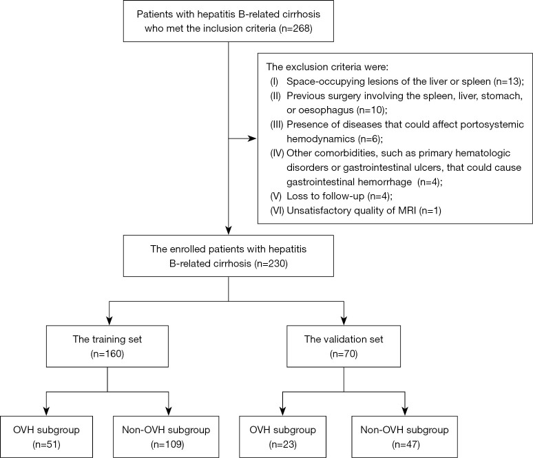
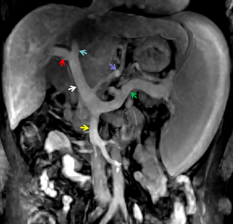
Methods: Two hundred and thirty consecutive individuals with hepatitis B-related cirrhosis undergoing abdominal enhanced MRI were randomly grouped into training (n=160) and validation sets (n=70). OVH was confirmed in 51 and 23 participants in the training and validation sets during 2-year follow-up period, respectively. Spleen, total liver, right lobe, caudate lobe, left lateral lobe, and left medial lobe volumes, together with diameters of maximum OV and portal venous system were measured on MRI. In the training set, univariate analyses and binary logistic regression analyses were conducted to determine independent predictors. The performance of the model for predicting OVH constructed based on independent predictors from the training set was evaluated with receiver operating characteristic (ROC) analysis and validated in the validation set.
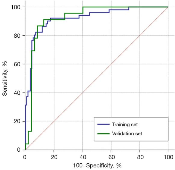
Results: The model for predicting OVH was established based on right liver lobe volume and diameters of the maximum OV, left gastric vein, and portal vein [odds ratio (OR) =0.991, 2.462, 1.434, and 1.582, respectively; all P values <0.05]. The logistic regression model equation [-0.009 × right liver lobe volume + 0.901 × maximum OV diameter (MOVD) + 0.361 × left gastric vein diameter (LGVD) + 0.459 × portal vein diameter (PVD) - 7.842] with a cutoff value of -0.656 for predicting OVH obtained excellent performance with an area under ROC curve (AUC) of 0.924 [95% confidence interval (CI): 0.878-0.971]. The Delong test showed negative statistical difference in the model performance between the training and validation sets, with a P value >0.99.
Conclusions: The model could help well screen those patients at high risk of OVH for timely intervention and avoiding the fatal complications.
Keywords: Gastrointestinal haemorrhage; liver cirrhosis; magnetic resonance imaging (MRI); portal vein.
2023 Quantitative Imaging in Medicine and Surgery. All rights reserved.
Conflict of interest statement
Conflicts of Interest: All authors have completed the ICMJE uniform disclosure form (available at https://qims.amegroups.com/article/view/10.21037/qims-23-353/coif). The authors have no conflicts of interest to declare.
Figures

Similar articles
-
A novel model based on liver/spleen volumes and portal vein diameter on MRI to predict variceal bleeding in HBV cirrhosis.Eur Radiol. 2023 Feb;33(2):1378-1387. doi: 10.1007/s00330-022-09107-5. Epub 2022 Sep 1. Eur Radiol. 2023. PMID: 36048206 Clinical Trial.
-
Gadobenate dimeglumine-enhanced magnetic resonance imaging can accurately predict the severity of esophageal varices and portal vein pressure in patients with hepatitis B cirrhosis.J Dig Dis. 2020 Feb;21(2):104-111. doi: 10.1111/1751-2980.12843. J Dig Dis. 2020. PMID: 31922658
-
Combinations of liver lobe and spleen volumes obtained on magnetic resonance imaging to predict esophagogastric variceal bleeding in hepatitis B-related cirrhotic patients: A prospective cohort study.Medicine (Baltimore). 2022 Sep 23;101(38):e30616. doi: 10.1097/MD.0000000000030616. Medicine (Baltimore). 2022. PMID: 36197258 Free PMC article.
-
Band ligation versus sham or no intervention for primary prophylaxis of oesophageal variceal bleeding in children and adolescents with chronic liver disease or portal vein thrombosis.Cochrane Database Syst Rev. 2021 Jan 26;1(1):CD011561. doi: 10.1002/14651858.CD011561.pub2. Cochrane Database Syst Rev. 2021. PMID: 33522602 Free PMC article.
-
Sclerotherapy versus sham or no intervention for primary prophylaxis of oesophageal variceal bleeding in children with chronic liver disease or portal vein thrombosis.Cochrane Database Syst Rev. 2020 Mar 5;3(3):CD011573. doi: 10.1002/14651858.CD011573.pub2. Cochrane Database Syst Rev. 2020. PMID: 32133620 Free PMC article.
Cited by
-
Continuous Non-Invasive Hemodynamic Monitoring in Cirrhotic Patients-Friend or Foe?Medicina (Kaunas). 2025 Mar 19;61(3):536. doi: 10.3390/medicina61030536. Medicina (Kaunas). 2025. PMID: 40142347 Free PMC article.
References
LinkOut - more resources
Full Text Sources