

Comparison of cardiovascular metrics on computed tomography pulmonary angiography of the updated and old diagnostic criteria for pulmonary hypertension in patients with chronic thromboembolic pulmonary hypertension
- PMID: 38106317
- PMCID: PMC10721984
- DOI: 10.21037/qims-23-250
Comparison of cardiovascular metrics on computed tomography pulmonary angiography of the updated and old diagnostic criteria for pulmonary hypertension in patients with chronic thromboembolic pulmonary hypertension
Abstract
Background: In the 2022 European Society of Cardiology (ESC) and the European Respiratory Society (ERS) guidelines, the diagnostic criteria for pulmonary hypertension (PH) included a reduced mean pulmonary artery pressure (mPAP) of 20 mmHg (mPAP >20 mmHg). This study aimed to reassess cardiovascular metrics on computed tomography pulmonary angiography (CTPA) for chronic thromboembolic pulmonary hypertension (CTEPH) to optimize the timely diagnosis of patients with suspected PH.
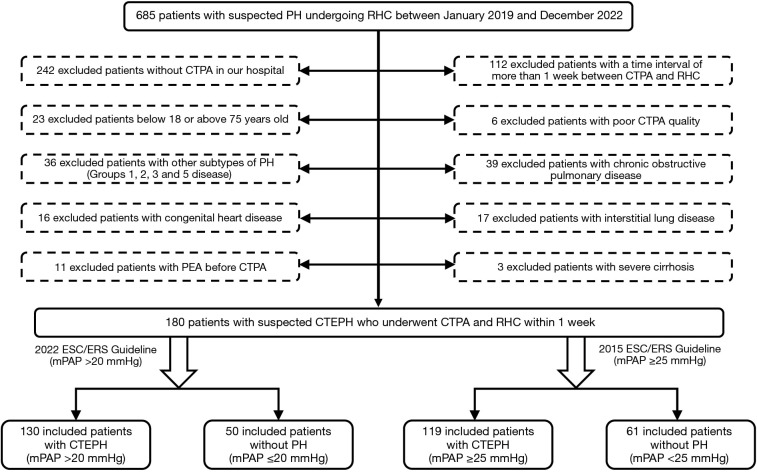
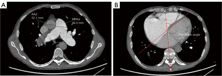
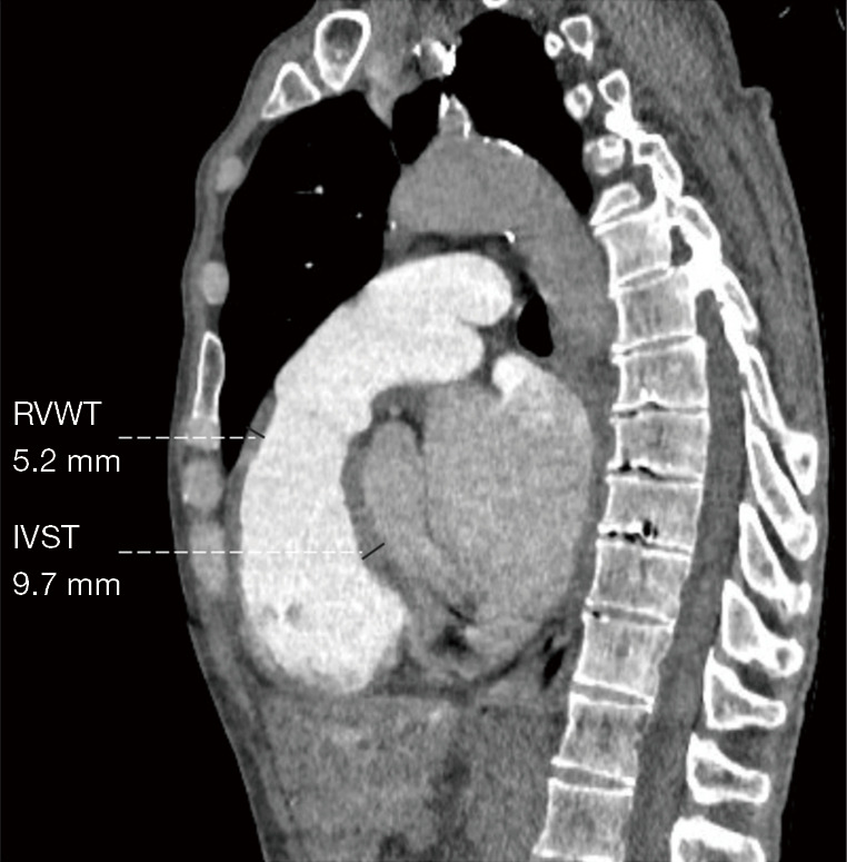
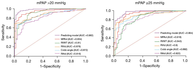
Methods: Patients with suspected CTEPH who underwent CTPA and right heart catheterization (RHC) between January 2019 and December 2022 in China-Japan Friendship Hospital were retrospectively included. They were grouped into CTEPH and non-PH groups according to the new and old criteria (2022 and 2015 ESC/ERS guidelines) for the diagnosis of PH. Cardiovascular metrics including the main pulmonary artery diameter (MPAd), Cobb angle, and right ventricular free wall thickness (RVWT), among others, were measured. The correlation of these metrics with hemodynamic data was analyzed with Spearman rank correlation analysis, while the differences in cardiovascular metrics between the updated (mPAP >20 mmHg) and old PH criteria (mPAP ≥25 mmHg) were compared with independent samples t-test or the Mann-Whitney test. Receiver operator characteristic (ROC) curve analysis was performed for the prediction model.
Results: The study enrolled 180 patients (males n=86; age 55.5±12.0 years old). According to the old guidelines, 119 patients were placed into the PH group (mPAP ≥25 mmHg) , while according to the new guidelines, 130 patients were placed into the PH group (mPAP >20 mmHg). Cardiovascular metrics on CTPA between the updated and old guidelines were comparable (P>0.05). Compared to other metrics, an MPAd of 30.4 mm exhibited the highest area under the curve (AUC: 0.934±0.021), with a sensitivity of 0.88 and specificity of 0.90. MPAd [odds ratio (OR) =1.271], transverse diameter of the right ventricle (RVtd; OR =1.176), Cobb angle (OR =1.108), and RVWT (OR =3.655) were independent factors for diagnosing CTEPH (P<0.05). Cobb angle, right and left ventricular transverse diameter ratio, and right and left ventricular area ratio moderately correlated with mPAP (r=0.586, r=0.583, r=0.629) and pulmonary vascular resistance (PVR) (r=0.613, r=0.593, r=0.642).
Conclusions: Cardiovascular metrics on CTPA were comparable between the new and old guidelines for CTEPH diagnosis. Cardiovascular metrics on CTPA can noninvasively assess the hemodynamics of patients with CTEPH.
Keywords: Pulmonary hypertension (PH); chronic thromboembolic pulmonary hypertension (CTEPH); computed tomography pulmonary angiography (CTPA); hemodynamics.
2023 Quantitative Imaging in Medicine and Surgery. All rights reserved.
Conflict of interest statement
Conflicts of Interest: All authors have completed the ICMJE uniform disclosure form (available at https://qims.amegroups.com/article/view/10.21037/qims-23-250/coif). The authors have no conflicts of interest to declare.
Figures

Similar articles
-
Cardiovascular metrics on CT pulmonary angiography in patients with pulmonary hypertension - re-evaluation under the updated guidelines of pulmonary hypertension.Insights Imaging. 2023 Oct 23;14(1):179. doi: 10.1186/s13244-023-01535-1. Insights Imaging. 2023. PMID: 37872384 Free PMC article.
-
Noninvasive prediction of pulmonary hemodynamics in chronic thromboembolic pulmonary hypertension by electrocardiogram-gated computed tomography.Eur J Radiol Open. 2021 Oct 18;8:100384. doi: 10.1016/j.ejro.2021.100384. eCollection 2021. Eur J Radiol Open. 2021. PMID: 34712746 Free PMC article.
-
Cardiovascular parameters of computed tomographic pulmonary angiography to assess pulmonary vascular resistance in patients with chronic thromboembolic pulmonary hypertension.Int J Cardiol. 2013 Apr 15;164(3):295-300. doi: 10.1016/j.ijcard.2011.07.019. Epub 2011 Aug 5. Int J Cardiol. 2013. PMID: 21820745
-
Chronic thromboembolic pulmonary hypertension (CTEPH) - potential role of multidetector-row CT (MD-CT) and MR imaging in the diagnosis and differential diagnosis of the disease.Rofo. 2014 Aug;186(8):751-61. doi: 10.1055/s-0034-1366425. Epub 2014 Apr 22. Rofo. 2014. PMID: 24756429 Review.
-
Chronic thromboembolic pulmonary hypertension.Presse Med. 2015 Dec;44(12 Pt 2):e409-16. doi: 10.1016/j.lpm.2015.10.010. Epub 2015 Nov 12. Presse Med. 2015. PMID: 26585271 Review.
References
-
- Humbert M, Kovacs G, Hoeper MM, Badagliacca R, Berger RMF, Brida M, Carlsen J, Coats AJS, Escribano-Subias P, Ferrari P, Ferreira DS, Ghofrani HA, Giannakoulas G, Kiely DG, Mayer E, Meszaros G, Nagavci B, Olsson KM, Pepke-Zaba J, Quint JK, Rådegran G, Simonneau G, Sitbon O, Tonia T, Toshner M, Vachiery JL, Vonk Noordegraaf A, Delcroix M, Rosenkranz S, ESC/ERS Scientific Document Group . 2022 ESC/ERS Guidelines for the diagnosis and treatment of pulmonary hypertension. Eur Heart J 2022;43:3618-731. Erratum in: Eur Heart J 2023;44:1312. - PubMed
-
- Galiè N, Humbert M, Vachiery JL, Gibbs S, Lang I, Torbicki A, Simonneau G, Peacock A, Vonk Noordegraaf A, Beghetti M, Ghofrani A, Gomez Sanchez MA, Hansmann G, Klepetko W, Lancellotti P, Matucci M, McDonagh T, Pierard LA, Trindade PT, Zompatori M, Hoeper M, ESC Scientific Document Group . 2015 ESC/ERS Guidelines for the diagnosis and treatment of pulmonary hypertension: The Joint Task Force for the Diagnosis and Treatment of Pulmonary Hypertension of the European Society of Cardiology (ESC) and the European Respiratory Society (ERS): Endorsed by: Association for European Paediatric and Congenital Cardiology (AEPC), International Society for Heart and Lung Transplantation (ISHLT). Eur Heart J 2016;37:67-119. 10.1183/13993003.01032-2015 - DOI - PubMed
-
- Klok FA, Barco S, Konstantinides SV, Dartevelle P, Fadel E, Jenkins D, Kim NH, Madani M, Matsubara H, Mayer E, Pepke-Zaba J, Delcroix M, Lang IM. Determinants of diagnostic delay in chronic thromboembolic pulmonary hypertension: results from the European CTEPH Registry. Eur Respir J 2018;52:1801687. 10.1183/13993003.01687-2018 - DOI - PubMed
-
- Swift AJ, Dwivedi K, Johns C, Garg P, Chin M, Currie BJ, Rothman AM, Capener D, Shahin Y, Elliot CA, Charalampopolous T, Sabroe I, Rajaram S, Hill C, Wild JM, Condliffe R, Kiely DG. Diagnostic accuracy of CT pulmonary angiography in suspected pulmonary hypertension. Eur Radiol 2020;30:4918-29. 10.1007/s00330-020-06846-1 - DOI - PMC - PubMed
LinkOut - more resources
Full Text Sources
Research Materials