

Discrepancy in the suppressive function of regulatory T cells in allergic asthmatic vs. allergic rhinitis subjects upon low-dose allergen challenges
- PMID: 38106504
- PMCID: PMC10722309
- DOI: 10.3389/falgy.2023.1296601
Discrepancy in the suppressive function of regulatory T cells in allergic asthmatic vs. allergic rhinitis subjects upon low-dose allergen challenges
Abstract
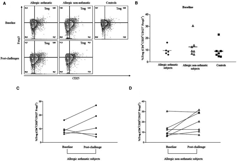
Background: Regulatory T cells (Tregs) contribute to the maintenance of immunological tolerance. There is evidence of impaired function of these cells in people with asthma and allergy. In this study, we evaluated and compared the function of Tregs in allergic asthmatic and allergic non-asthmatic patients, both before and after low-dose allergen challenges.
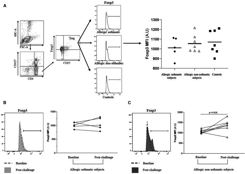
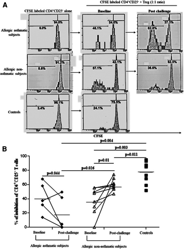
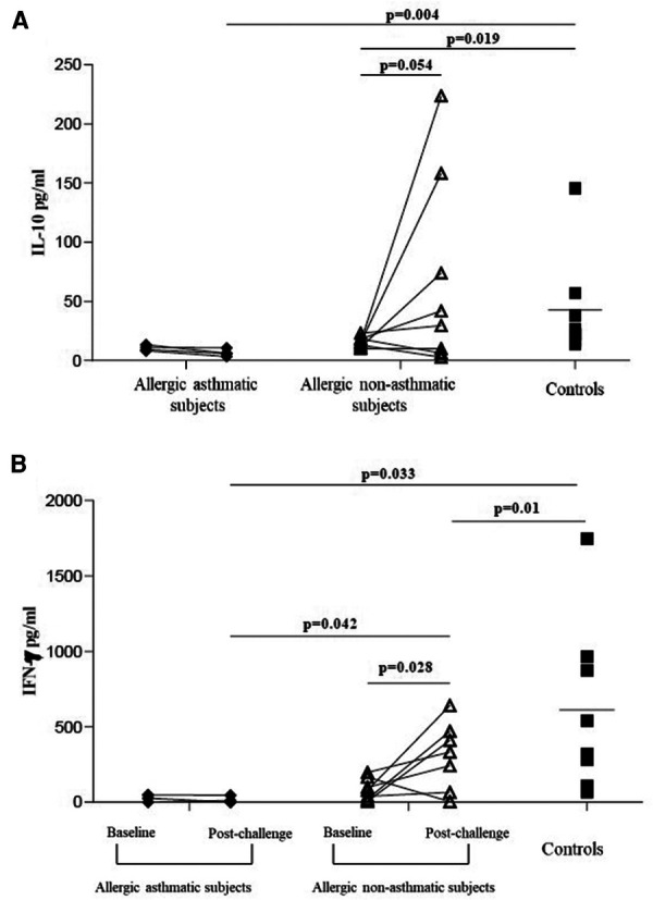
Methods: Three groups of subjects were recruited for a baseline evaluation: healthy controls without allergy or asthma, allergic asthmatic subjects, and allergic non-asthmatic subjects. All of them were subjected to expiratory flow measurements, sputum induction, and blood sampling. In addition, both groups of allergic subjects underwent low-dose allergen challenges. Tregs were isolated from whole blood using CD4+CD25high and CD127low staining. The suppression function was measured by flow cytometry. The levels of IL-10, IFN-γ, IgG4, IgA, and TGF-β were measured using ELISA, and sputum Foxp3 was evaluated using qRT-PCR.
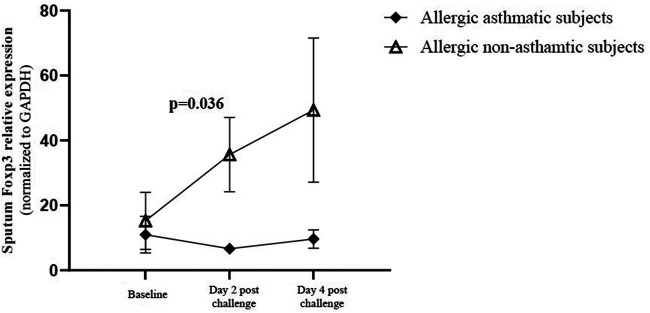
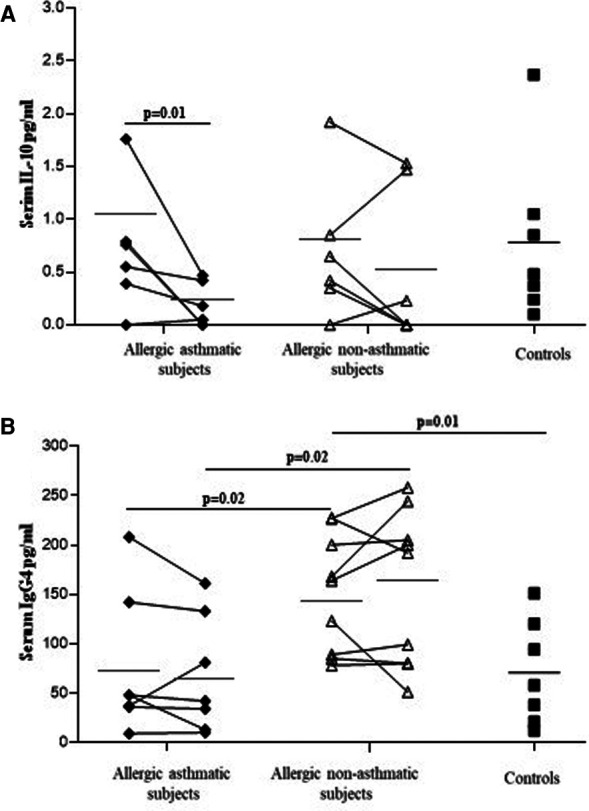
Results: The suppressive function of Tregs in healthy controls was significantly higher than in allergic asthmatic or allergic non-asthmatic subjects. Repeated exposure to low doses of allergen increased the suppressor function of Tregs in allergic non-asthmatic subjects but decreased it in allergic asthmatic subjects. Foxp3 gene expression was increased in induced sputum in allergic non-asthmatic subjects, whereas it did not change in asthmatic subjects. Serum IL-10 level was decreased in allergic asthmatic subjects after allergen challenge but not in allergic non-asthmatic subjects. IFN-γ level increased upon allergen challenge in allergic non-asthmatic subjects. IgG4 level was higher in allergic non-asthmatic subjects than in allergic asthmatic subjects.
Conclusions: Low-dose allergen challenges stimulate the suppressor function of Tregs in non-asthmatic allergic subjects but not in allergic asthmatic subjects.
Keywords: Foxp3; IL-10; allergen exposure; allergy; bronchoalveolar lavage fluid (BALF), Tregs.
© 2023 Klein, Plante, Boulay, Boulet and Chakir.
Conflict of interest statement
The authors declare that the research was conducted in the absence of any commercial or financial relationships that could be construed as a potential conflict of interest.
Figures
Similar articles
-
Allergen provocation increases TH2-cytokines and FOXP3 expression in the asthmatic lung.Allergy. 2010 Mar;65(3):311-8. doi: 10.1111/j.1398-9995.2009.02218.x. Epub 2009 Oct 20. Allergy. 2010. PMID: 19845574
-
Allergen-induced bronchial inflammation in house dust mite-allergic patients with or without asthma.Clin Exp Allergy. 2002 Dec;32(12):1720-7. doi: 10.1046/j.1365-2222.2002.01542.x. Clin Exp Allergy. 2002. PMID: 12653162
-
A single nasal allergen challenge increases induced sputum inflammatory markers in non-asthmatic subjects with seasonal allergic rhinitis: correlation with plasma interleukin-5.Clin Exp Allergy. 2003 Apr;33(4):475-82. doi: 10.1046/j.1365-2222.2003.01632.x. Clin Exp Allergy. 2003. PMID: 12680863 Clinical Trial.
-
Insufficient increment of CD4+CD25+ regulatory T cells after stimulation in vitro with allergen in allergic asthma.Int Arch Allergy Immunol. 2009;148(3):199-210. doi: 10.1159/000161580. Epub 2008 Oct 10. Int Arch Allergy Immunol. 2009. PMID: 18849611
-
Foxp3 expressing regulatory T-cells in allergic disease.Adv Exp Med Biol. 2009;665:180-94. doi: 10.1007/978-1-4419-1599-3_14. Adv Exp Med Biol. 2009. PMID: 20429425 Review.
References
LinkOut - more resources
Full Text Sources
Research Materials
Miscellaneous