. 2023 Oct 10:65:102253.
doi: 10.1016/j.eclinm.2023.102253. eCollection 2023 Nov.

Development of neonatal-specific sequences for portable ultralow field magnetic resonance brain imaging: a prospective, single-centre, cohort study

Collaborators, Affiliations

Development of neonatal-specific sequences for portable ultralow field magnetic resonance brain imaging: a prospective, single-centre, cohort study

Paul Cawley et al. EClinicalMedicine. .

Abstract

Background: Magnetic Resonance (MR) imaging is key for investigation of suspected newborn brain abnormalities. Access is limited in low-resource settings and challenging in infants needing intensive care. Portable ultralow field (ULF) MRI is showing promise in bedside adult brain imaging. Use in infants and children has been limited as brain-tissue composition differences necessitate sequence modification. The aim of this study was to develop neonatal-specific ULF structural sequences and test these across a range of gestational maturities and pathologies to inform future validation studies.

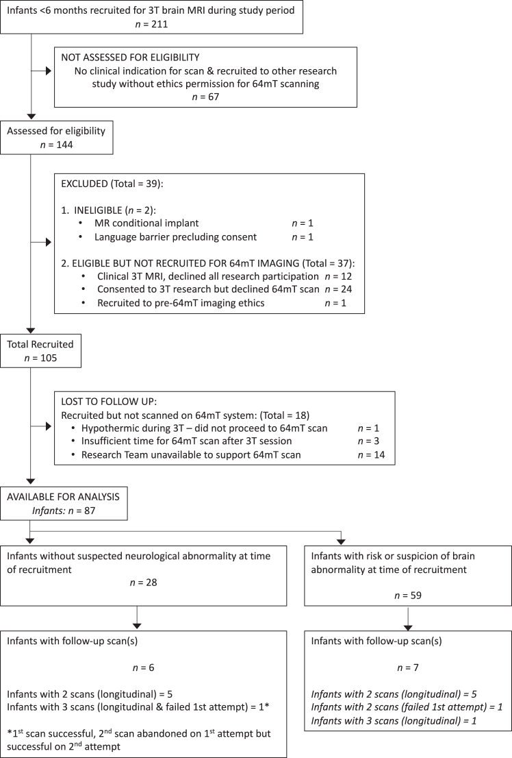
Methods: Prospective cohort study within a UK neonatal specialist referral centre. Infants undergoing 3T MRI were recruited for paired ULF (64mT) portable MRI by convenience sampling from the neonatal unit and post-natal ward. Key inclusion criteria: 1) Infants with risk or suspicion of brain abnormality, or 2) preterm and term infants without suspicion of major genetic, chromosomal or neurological abnormality. Exclusions: presence of contra-indication for MR scanning. ULF sequence parameters were optimised for neonatal brain-tissues by iterative and explorative design. Neuroanatomic and pathologic features were compared by unblinded review, informing optimisation of subsequent sequence generations in a step-wise manner. Main outcome: visual identification of healthy and abnormal brain tissues/structures. ULF MR spectroscopy, diffusion, susceptibility weighted imaging, arteriography, and venography require pre-clinical technical development and have not been tested.

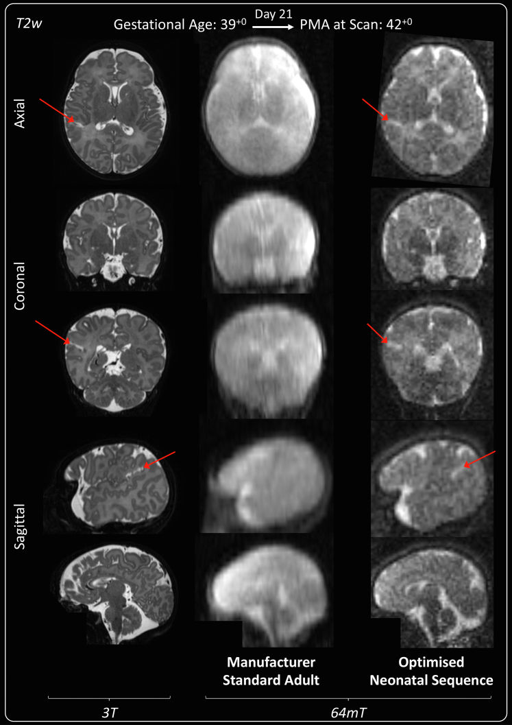
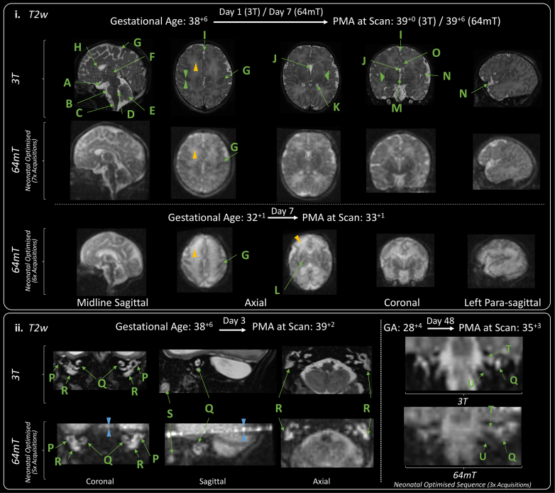
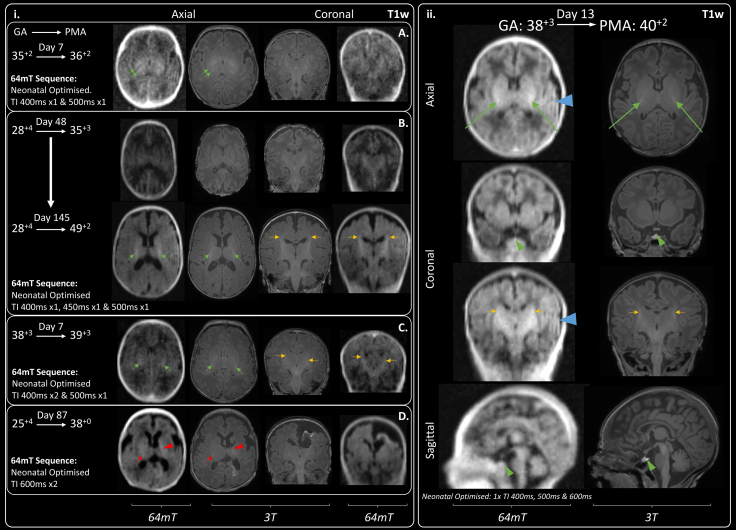
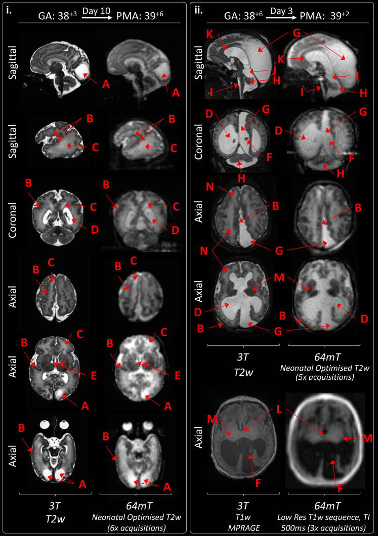
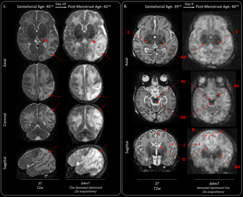
Findings: Between September 23, 2021 and October 25, 2022, 102 paired scans were acquired in 87 infants; 1.17 paired scans per infant. Median age 9 days, median postmenstrual age 40+2 weeks (range: 31+3-53+4). Infants had a range of intensive care requirements. No adverse events observed. Optimised ULF sequences can visualise key neuroanatomy and brain abnormalities. In finalised neonatal sequences: T2w imaging distinguished grey and white matter (7/7 infants), ventricles (7/7), pituitary tissue (5/7), corpus callosum (7/7) and optic nerves (7/7). Signal congruence was seen within the posterior limb of the internal capsule in 10/11 infants on finalised T1w scans. In addition, brain abnormalities visualised on ULF optimised sequences have similar MR signal patterns to 3T imaging, including injury secondary to infarction (6/6 infants on T2w scans), hypoxia-ischaemia (abnormal signal in basal ganglia, thalami and white matter 2/2 infants on T2w scans, cortical highlighting 1/1 infant on T1w scan), and congenital malformations: polymicrogyria 3/3, absent corpus callosum 2/2, and vermian hypoplasia 3/3 infants on T2w scans. Sequences are susceptible to motion corruption, noise, and ULF artefact. Non-identified pathologies were small or subtle.

Interpretation: On unblinded review, optimised portable MR can provide sufficient contrast, signal, and resolution for neuroanatomical identification and detection of a range of clinically important abnormalities. Blinded validation studies are now warranted.

Funding: The Bill and Melinda Gates Foundation, the MRC, the Wellcome/EPSRC Centre for Medical Engineering, the MRC Centre for Neurodevelopmental Disorders, and the National Institute for Health Research (NIHR) Biomedical Research Centres based at Guy's and St Thomas' and South London & Maudsley NHS Foundation Trusts and King's College London.

Keywords: Intensive care; Low field; Magnetic resonance imaging; Neonatal; Portable.


Conflict of interest statement

PC is supported by the Medical Research Council Centre for Neurodevelopmental Disorders [MR/N026063/1] to undertake this work and received an educational stipend from ISMRM to attend the international ISMRM conference and present this work. FP was employed by the Guy′s & St. Thomas′ NHS Foundation Trust & King’s College London as a senior MR physicist at study outset, during experimental design and initial participant recruitment, he is now a senior clinical scientist employed by Hyperfine Inc., since May 2022–drawing a salary, shares and stock options. RT is an MR sequence developer employed by Hyperfine Inc., and is a holder of shares and stock option of Hyperfine Inc. JOM has institutional funding from the Bill & Melinda Gates Foundation Consortium grant to support research work by his group in neurodevelopment, including data from conventional 3T MRI and Hyperfine scanners–this grant is focused on image analysis and is not commercially sponsored. SW has received funding from the Bill & Melinda Gates Foundation for attendance and travel to sites for training and knowledge exchange during the development and delivery of this project. TA is supported by the UK MRC for a Translational support fellowship [MR/V036874/1] for personal salary, and funding for 3T MRI scans, the MRC Centre for Neurodevelopmental Disorders, King’s College London [MR/N026063/1] for administrative support, funding, and a Clinician Scientist Fellowship [MR/P008712/1]—for personal salary and funding for 3T MRI scans. TA is supported by the EPSRC UK Network grant, co-investigator [EP/W035154/1]. DE and JVH: The Hyperfine machine (Swoop® MR System) was provided by the Bill and Melinda Gates Foundation as part of the Unity Consortium.

Figures

Fig. 1
Fig. 1
Patient recruitment flow diagram. 102 scans (including 2× failed attempts) in 87 participants.
Fig. 2
Fig. 2
T2 weighted manufacturer standard adult 64mT sequence compared with reference 3T imaging and dedicated 64mT neonatal sequence. Term infant with small right-sided focal cortical infarction in the superior parietal lobe (red arrows). 3T MR scan was performed on day 15 and 64mT scan on day 21. 64mT adult sequence shows poor tissue contrast, the cortical infarct is not identifiable and there is asymmetric signal inhomogeneity (generalised left sided high T2). Subtle abnormal cortical signal is present on the optimised 64mT neonatal sequence in the location of the infarct. Deep grey matter on axial plane and cerebellum on coronal plane remain unclear and PLIC not identified due to persistent noise and narrow contrast. PMA: post-menstrual age; PLIC: posterior limb of the internal capsule.
Fig. 3
Fig. 3
Neuroanatomy and structural imaging. (i) Example T2w imaging of the normally developed term and preterm brain. Green Double Arrowheads—Cortex. Green Single Wide Arrowheads—Deep Grey Matter. Amber Single Narrow Arrowhead—white matter (higher T2 signal is apparent in the preterm infant). Deep Grey Matter and medial temporal lobes appear as lower T2 intensity in comparison with 3T imaging. Labelled structures are visible on both 3T and 64mT imaging: A) Optic Nerve, B) Pons, C) Medulla, D) Fourth Ventricle, E) Cerebellar Vermis, F) Thalamus, G) Central Sulcus, H) Anterior Corpus Callosum, I) Interhemispheric Fissure, J) Cavum Septum Pellucidum, K) Lateral Ventricle, L) Internal Capsule (Only visible in the preterm infant imaging; as high T2 streak), M) Third Ventricle, N) Sylvian Fissure, O) Cingulate Gyrus. (ii) Inner Ear Structures. Zipper artefact (Blue double arrowheads) transects the inner ear structures in some infants. As shown, inner ear structures and cranial nerves can be difficult to delineate on standard high field (3T) acquisitions, without dedicated views. P) Semi-Circular Canal, Q) Cochlear, R) Vestibule, S) Tooth; and courses of the T) Vestibulocochlear Nerve, and U) Glossopharyngeal Nerve. GA: gestational age; PMA: post-menstrual age.
Fig. 4
Fig. 4
T1 signal within the Posterior Limb of the Internal Capsule (PLIC). Single green arrows indicate normal T1 hyperintensity within the PLIC and single amber arrows indicate normal T1 hyperintensity within the corticospinal tracts. (i) Case series demonstrating progression of T1 signal generated by myelination across range of gestations: A) Preterm infant with absence of myelin signal within the PLIC which appears hypointense due to normal for age high T1 signal within the ventrolateral nucleus posteriorly and globus pallidus anteriorly (double green arrows). B) Very preterm infant imaged during prematurity and again at post-term -demonstrating evolution of myelination of the full length of the PLIC by 49+2 weeks gestation. 64mT scan resolution may preclude visualisation of thin strip of T1 hyperintensity seen on 3T images within the anterior limb of the internal capsule. C) Small amount of T1 myelin signal seen within PLIC bilaterally, infant imaged just prior to their expected delivery date. D) Asymmetry in PLIC signal: small amount of T1 myelin signal on right-side (single red arrow), with abnormal high T1 signal for gestation along the left PLIC (red arrowhead) secondary to extensive left-sided haemorrhagic infarction. (ii) Exemplar 64mT T1w neonatal imaging—multi-inversion time sequence combination facilitates superimposition of PLIC signal alongside white matter, grey matter, and CSF contrast. Pituitary tissue is labelled with a green arrowhead. Ghosting artefact is evident on left-side (blue arrowhead), this requires further investigation. GA: gestational age; PMA: post-menstrual age.
Fig. 5
Fig. 5
Congenital structural anomalies and disrupted development. (i) In utero CMV infection leading large bilateral sub-occipital cysts (A), extensive polymicrogyria (some examples are given—B—depicted on 64mT as thickened cortex with shallow sulci), patchy WM T2 hyperintensities (C), ventriculomegaly (D), and bilateral subependymal cysts (E). (ii) Cerebellopontine Hypoplasia with severe ventriculomegaly (D) and malpositioned choroid plexi (F), leading to enlargement of a midline crossing inter-hemispheric cyst (G). The left cerebral hemisphere is smaller than the right, there is extensive polymicrogyria (examples given—B), hypoplastic cerebellar hemispheres and vermis (H), hypoplastic pons and brainstem (I), dilated quadrigeminal cistern (J), thin stretched corpus callosum (K), small deep grey matter structures with wide thalamic adhesion (L) and absence of myelin which is abnormal for this infant’s gestational maturity (M). White matter striations are seen on 3T T2w imaging (N)—some correlative findings are present on 64mT imaging, but these are ill-defined, presenting mainly as generalised high T2 white matter at ULF. Choroid plexi are markedly less distinct, but remain identifiable, on 64mT imaging. GA: gestational age; PMA: post-menstrual age; Low Res: low resolution.
Fig. 6
Fig. 6
Acquired injury in the normally formed brain. (i) Diffuse left sided white matter and cortical parieto-occipital infarction (red arrows). High T2 signal is also seen within the left posterior thalamus, suggestive of secondary network injury (red arrowhead). A video of this study is available as an Online Supplement. (ii) Bilateral basal ganglia, thalamic, cortico-spinal tract, and mesencephalon injury secondary to perinatal hypoxia-ischaemia. Hypoxic-ischaemic injury revealed as abnormal low T2 within the deep grey matter (Ad), cerebral peduncles (Ac), hippocampi (Ah) and brainstem (Ab). There is diffuse abnormal high T2 signal in the white matter (C), with areas of sub-cortical white matter highlighting on 64mT T2w images (D). The Posterior Limb of the Internal Capsule is abnormally T2 hyperintense -abnormal for gestational maturity (E). Additional findings include widened interhemispheric fissure (F), and bilateral sub-ependymal cysts (G).

References

    1. Montagna A., Karolis V., Batalle D., et al. ADHD symptoms and their neurodevelopmental correlates in children born very preterm. PLoS One. 2020;15 - PMC - PubMed
    1. Arulkumaran S., Tusor N., Chew A., et al. MRI findings at term-corrected age and neurodevelopmental outcomes in a large cohort of very preterm infants. Am J Neuroradiol. 2020;41:1509–1516. - PMC - PubMed
    1. Pappas A., Shankaran S., McDonald S.A., et al. Cognitive outcomes after neonatal encephalopathy. Pediatrics. 2015;135:e624–e634. - PMC - PubMed
    1. Schreglmann M., Ground A., Vollmer B., Johnson M.J. Systematic review: long-term cognitive and behavioural outcomes of neonatal hypoxic–ischaemic encephalopathy in children without cerebral palsy. Acta Paediatr. 2020;109:20–30. - PubMed
    1. Tonks J., Cloke G., Lee-Kelland R., et al. Attention and visuo-spatial function in children without cerebral palsy who were cooled for neonatal encephalopathy: a case-control study. Brain Inj. 2019;33:894–898. - PubMed

LinkOut - more resources