. 2023 Sep 4;34(1):150-153.
doi: 10.1055/s-0043-1772691. eCollection 2024 Jan.

Dorsal Wrist Carpal Boss Impingement-Dynamic Ultrasound to the Rescue!

Affiliations

Dorsal Wrist Carpal Boss Impingement-Dynamic Ultrasound to the Rescue!

Aakanksha Agarwal et al. Indian J Radiol Imaging. .

Abstract

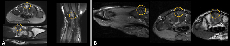
The "carpal boss" is a variant present in 19% of the population according to cadaveric studies but becomes symptomatic in only 1% of cases. With the rising popularity of "yoga," which includes prolonged hyperextension at the wrist joint with weight bearing, an increasing number of individuals with silent carpal boss present with dorsal wrist pain due to impingement over the dorsal soft tissues by this innocuous bony protuberance. This warrants the attention of radiologists and clinicians while dealing with wrist pain. It can be challenging to identify this bossing on routine radiographs, necessitating special views. We describe the use of dynamic ultrasound in diagnosing "symptomatic" carpal boss, the effects of which become even more evident on imaging during hyperextension-the triggering movement.

Keywords: carpal boss; dynamic ultrasound; impingement.


Conflict of interest statement

Conflict of Interest None declared.

Figures

Fig. 1
Fig. 1
( A ) Bony protuberance at the quadrangular joint at the dorsal aspect of the base of third metacarpal and the capitate (yellow arrow and circle). Extensor carpi radialis and digital extensor tendons in close proximity to the carpal boss are also depicted. ( B ) Increased dorsal soft tissue compartment pressure results in extrusion of a small ganglion (blue) with underlying synovial hypertrophy. Overlying carpal boss and traversing extensor tendons (purple).
Fig. 2
Fig. 2
( A ) a: Carpal bossing at the base of third metacarpal with dorsal osteophyte arising from capitate with bony remodeling (encircled). b: Carpal bossing at the base of third metacarpal with bony remodeling and cystic change at the carpometacarpal joint (encircled). c: Carpal bossing at the base of third metacarpal with bony remodeling and degenerative change at the dorsal aspect of the quadrangular joint (encircled). ( B ) Single-photon emission computed tomography demonstrating an unfused os styloideum with radioisotope avidity at the third carpometacarpal joint suggesting on going mechanical stress (encircled).
Fig. 3
Fig. 3
( A ) Magnetic resonance imaging proton density fat-saturated axial, sagittal T1, and PDFS coronal images demonstrating an unfused and fragmented os styloideum with prominent edema and cystic change at the synchondrosis at the third carpometacarpal joint suggesting stress (encircled). ( B ) Magnetic resonance proton density fat-saturated sagittal, axial, and T1 axial images demonstrating an unfused os styloideum with mild edema at the synchondrosis at the third carpometacarpal joint suggesting stress (encircled). GRE; gradient echo; PDFS, proton density fat saturated.
Fig. 4
Fig. 4
( A ) Dorsal soft tissue impingement with wrist hyperextension secondary to an underlying carpal boss (yellow), resulting in extrusion of hypertrophied synovium and small effusion (blue). ( B ) Magnetic resonance imaging sagittal T1 fat-saturated post-contrast image, axial T1, and coronal GRE images: Dorsal soft tissue impingement secondary to an underlying carpal boss (yellow), resulting in post-contrast enhancement of hypertrophied synovium and small effusion (blue). The patient-placed skin markers are along the third ray and correspond to the site of the bony protuberance (green arrows).
Fig. 5
Fig. 5
( A ) Snapping of third digit extensor tendon (red and blue) over a carpal boss (yellow) with finger extension. Associated small dorsal wrist ganglion. The probe position has been depicted in the inset. ( B ) Accessory extensor digitorum manus brevis (red) over carpal boss (yellow) resulting in dynamic impingement and dorsal wrist pain. The probe position has been depicted in the inset. ( C ) Splaying of second- and third-digit extensor tendons (red and green) around a dorsal wrist ganglion (blue) and underlying carpal boss (yellow) with finger extension. The probe position has been depicted in the inset. ( D ) Splaying of second- and third-digit extensor tendons (red and green) over dorsal wrist ganglion (blue) and underlying carpal boss (yellow) with finger extension.
Fig. 6
Fig. 6
Management options for symptomatic carpal boss—ultrasound-guided steroid injection and surgical excision.

References

    1. Vieweg H, Radmer S, Fresow R et al.Diagnosis and treatment of symptomatic carpal bossing. J Clin Diagn Res. 2015;9(10):RC01–RC03. - PMC - PubMed
    1. Porrino J, Maloney E, Chew F S. Current concepts of the carpal boss: pathophysiology, symptoms, clinical or imaging diagnosis, and management. Curr Probl Diagn Radiol. 2015;44(05):462–468. - PubMed
    1. Mespreuve M, Waked K, Verstraete K. Imaging findings at the quadrangular joint in carpal boss. J Belg Soc Radiol. 2017;101(01):21. - PMC - PubMed
    1. Arend C F. The carpal boss: a review of different sonographic findings. Radiol Bras. 2014;47(02):112–114. - PMC - PubMed
    1. Clarke A M, Wheen D J, Visvanathan S, Herbert T J, Conolly W B. The symptomatic carpal boss. Is simple excision enough? J Hand Surg [Br] 1999;24(05):591–595. - PubMed