. 2023 Oct 19;14(12):2731-2737.
doi: 10.1039/d3md00439b. eCollection 2023 Dec 13.

A covalent fragment-based strategy targeting a novel cysteine to inhibit activity of mutant EGFR kinase

Affiliations

A covalent fragment-based strategy targeting a novel cysteine to inhibit activity of mutant EGFR kinase

Naoki Kuki et al. RSC Med Chem. .

Abstract

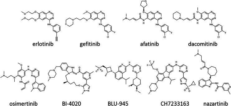
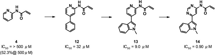
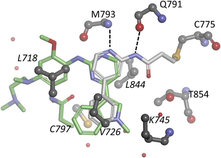
Several generations of ATP-competitive anti-cancer drugs that inhibit the activity of the intracellular kinase domain of the epidermal growth factor receptor (EGFR) have been developed over the past twenty years. The first-generation of drugs such as gefitinib bind reversibly and were followed by a second-generation such as dacomitinib that harbor an acrylamide moiety that forms a covalent bond with C797 in the ATP binding pocket. Resistance emerges through mutation of the T790 gatekeeper residue to methionine, which introduces steric hindrance to drug binding and increases the Km for ATP. A third generation of drugs, such as osimertinib were developed which were effective against T790M EGFR in which an acrylamide moiety forms a covalent bond with C797, although resistance has emerged by mutation to S797. A fragment-based screen to identify new starting points for an EGFR inhibitor serendipitously identified a fragment that reacted with C775, a previously unexploited residue in the ATP binding pocket for a covalent inhibitor to target. A number of acrylamide containing fragments were identified that selectively reacted with C775. One of these acrylamides was optimized to a highly selective inhibitor with sub-1 μM activity, that is active against T790M, C797S mutant EGFR independent of ATP concentration, providing a potential new strategy for pan-EGFR mutant inhibition.


Conflict of interest statement

There is no conflict of interest to declare.

Figures

Fig. 1
Fig. 1. Representative EGFR inhibitors.
Fig. 2
Fig. 2. The structure of osimertinib (stick, pink carbons) bound to EGFR_wtX (PDB code 6JXT) (schematic in white) with the side chain of residues that are mutated in various forms at positions 775, 790, 797, 858, 865, 866 and 867 in Corey–Pauling–Koltun model (CPK). Note that in this crystal structure C775 has two alternate conformers modeled (only conformer A shown) and there are several missing loops, including A865.
Fig. 3
Fig. 3. Detail from the crystal structure (PDB code: 8HV1) of fragment 1 (in stick with grey carbon atoms) bound to EGFR_DMX. Water molecules and labelled amino acids in ball and stick (grey carbon atoms) with main chain only for Q791 and M793, and hydrogen bonds in dashed lines. Details of all crystal structure determination in this paper are in ESI with data collection (Table S1†) and refinement statistics (Table S6†).
Fig. 4
Fig. 4. Acrylamide containing compounds selected from docking to the structure of EGFR_DMX.
Fig. 5
Fig. 5. Details from the crystal structures of EGFR_TMX with bound acrylamide compounds; (legend as Fig. 3) with carbon atoms for A. fragment 4 in light blue (PDB code: 8HV4); B. fragment 8 in green (PDB code 8HV6) and C. fragment 10 in orange (PDB code: 8HV8).
Fig. 6
Fig. 6. Optimization of acrylamide 4; IC50 is concentration of compound giving 50% inhibition of activity after incubation for 1.5 hours.
Fig. 7
Fig. 7. Details from the crystal structure of EGFR_TMX with bound 12 (PDB code: 8HV9, white carbon atoms, legend as in Fig. 3) with the structure of osimertinib with lime green carbons as bound to EGFR_wtX (PDB: 6JXT) showing the side chains of additional selected amino acids labelled in italics.
Fig. 8
Fig. 8. Detail of the crystal structure of 14 (magenta carbon atoms) bound to EGFR_TMX (PDB code: 8HV10); legend as in Fig. 3 with the side chain atoms of V726, L718, V726, K746 and L844 also shown.

References

    1. Cohen S. The stimulation of epidermal proliferation by a specific protein (EGF) Dev. Biol. 1965;12:394–407. doi: 10.1016/0012-1606(65)90005-9. - DOI - PubMed
    1. Wee P. Wang Z. Epidermal Growth Factor Receptor Cell Proliferation Signaling Pathways. Cancers. 2017;9(5):52. doi: 10.3390/cancers9050052. - DOI - PMC - PubMed
    1. Das D. Wang J. Hong J. Next-Generation Kinase Inhibitors Targeting Specific Biomarkers in Non-Small Cell Lung Cancer (NSCLC): A Recent Overview. ChemMedChem. 2021;16:2459–2479. doi: 10.1002/cmdc.202100166. - DOI - PubMed
    1. Lee C.-S. Sharma S. Miao E. Mensah C. Sullivan K. Seetharamu N. A Comprehensive Review of Contemporary Literature for Epidermal Growth Factor Receptor Tyrosine Kinase Inhibitors in Non-Small Cell Lung Cancer and Their Toxicity. Lung Cancer. 2020;11:73–103. - PMC - PubMed
    1. Grabe T. Lategahn J. Rauh D. C797S Resistance: The Undruggable EGFR Mutation in Non-Small Cell Lung Cancer? ACS Med. Chem. Lett. 2018;9(8):779–782. doi: 10.1021/acsmedchemlett.8b00314. - DOI - PMC - PubMed

LinkOut - more resources