. 2023 Jun 30;11(6):10.18103/mra.v11i6.3994.
doi: 10.18103/mra.v11i6.3994. Epub 2023 Jun 26.

The Glioblastoma Landscape: Hallmarks of Disease, Therapeutic Resistance, and Treatment Opportunities

Affiliations

The Glioblastoma Landscape: Hallmarks of Disease, Therapeutic Resistance, and Treatment Opportunities

Jack Boylan et al. Med Res Arch. .

Abstract

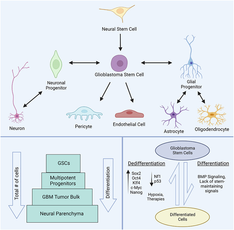
Malignant brain tumors are aggressive and difficult to treat. Glioblastoma is the most common and lethal form of primary brain tumor, often found in patients with no genetic predisposition. The median life expectancy for individuals diagnosed with this condition is 6 months to 2 years and there is no known cure. New paradigms in cancer biology implicate a small subset of tumor cells in initiating and sustaining these incurable brain tumors. Here, we discuss the heterogenous nature of glioblastoma and theories behind its capacity for therapy resistance and recurrence. Within the cancer landscape, cancer stem cells are thought to be both tumor initiators and major contributors to tumor heterogeneity and therapy evasion and such cells have been identified in glioblastoma. At the cellular level, disruptions in the delicate balance between differentiation and self-renewal spur transformation and support tumor growth. While rapidly dividing cells are more sensitive to elimination by traditional treatments, glioblastoma stem cells evade these measures through slow division and reversible exit from the cell cycle. At the molecular level, glioblastoma tumor cells exploit several signaling pathways to evade conventional therapies through improved DNA repair mechanisms and a flexible state of senescence. We examine these common evasion techniques while discussing potential molecular approaches to better target these deadly tumors. Equally important, the presented information encourages the idea of augmenting conventional treatments with novel glioblastoma stem cell-directed therapies, as eliminating these harmful progenitors holds great potential to modulate tumor recurrence.

Keywords: Glioblastoma; Glioblastoma stem cells; clinical treatments; heterogeneity; therapy resistance; tumor microenvironment.


Conflict of interest statement

Conflict of interest statement. The authors have no conflicts of interest to declare.

Figures

Figure 1.
Figure 1.. Tumor heterogeneity within glioblastoma lesions.
Tumor heterogeneity can be explained by the cancer stem cell (CSC) theory. In this hierarchical model, a single transformed stem cell (purple) can self-renew to create another CSC and a rapidly cycling multipotent progenitor (blue/green). These progenitors divide to supply the tumor bulk with differentiated cells (red). Heterogeneity arises from the degrees of differentiation through the tumor. The clonal evolution theory can also be incorporated into this framework through mutational events during CSC self-renewal to create a genetically novel CSC (orange) or by dedifferentiation of a differentiated cell back into a CSC (pink). These populations of CSCs and their descendants can then compete through natural selection of growth advantages. Created with BioRender.
Figure 2.
Figure 2.. Organization and development of glioblastoma stem cells.
Glioblastoma stem cells (GSCs) are multipotent and capable of differentiation into multiple cell lineages including both neuronal and glial cells. Pericytes and endothelial cells can also be differentiated from GSCs to form the expanding vasculature in GBM tumors. Glioblastoma stem cells can originate from transformed neural stem cells or from dedifferentiation of differentiated brain cells (top panel). Representation of the sub-populations of GSCs relative to other tumor cell constituents and differentiation status (bottom left panel). The conversion between GSCs and differentiated cells within tumors is regulated by a variety of signaling molecules, oncogenes, and environmental conditions (bottom right panel). Created with BioRender.
Figure 3.
Figure 3.. Niches that define the glioblastoma tumor microenvironment.
Defined regions within the tumor microenvironment are contained within specific niches. Glioblastoma tumors contain three major niches including the perivascular niche (left), the hypoxic niche (center), and the invasive niche (right). A variety of cell types and populations comprising each niche are depicted in the top panels. Corresponding histological features of niche components are displayed below each schematic. Denoted are: a glomeruloid microvascular proliferation (bottom left), pseudopalisading cells circumscribing necrosis (bottom center), leading tumor edge (bottom right). Histology images adapted from online resources-. Created with BioRender.
Figure 4.
Figure 4.. Therapy evasion of glioblastoma cells through senescence.
Glioblastoma cells (top left) that resist radio/chemotherapy are enriched for therapy-resistant mesenchymal (MES) signatures. Some assume therapy induced senescence, bypassing normal apoptosis processes. Senescent tumor cells (top middle) over time can recur to give rise to new masses (top right). During senescence, cells secrete cytokines, chemokines, matrix metalloproteinases, and other growth factors known as the senescence-associated secretory phenotype (SASP). These collective signals support stem maintenance by activating the Wnt pathway. Factors from the SASP can also contribute to invasion, migration, and angiogenesis supporting tumor recurrence. Created with BioRender.

References

    1. Rock K, Mcardle O, Forde P, et al. A clinical review of treatment outcomes in glioblastoma multiforme—the validation in a non-trial population of the results of a randomised Phase III clinical trial: has a more radical approach improved survival? Br J Radiol. 2012;85(1017):e729–e733. doi:10.1259/bjr/83796755 - DOI - PMC - PubMed
    1. Hanif F, Muzaffar K, Perveen K, Malhi SM, Simjee SU. Glioblastoma Multiforme: A Review of its Epidemiology and Pathogenesis through Clinical Presentation and Treatment. Asian Pac J Cancer Prev APJCP. 2017;18(1):3–9. doi:10.22034/APJCP.2017.18.1.3 - DOI - PMC - PubMed
    1. Ostrom QT, Gittleman H, Liao P, et al. CBTRUS Statistical Report: Primary Brain and Central Nervous System Tumors Diagnosed in the United States in 2007–2011. Neuro-Oncol. 2014;16(suppl_4):iv1–iv63. doi:10.1093/neuonc/nou223 - DOI - PMC - PubMed
    1. Stupp R, Mason WP, van den Bent MJ, et al. Radiotherapy plus Concomitant and Adjuvant Temozolomide for Glioblastoma. N Engl J Med. 2005;352(10):987–996. doi:10.1056/NEJMoa043330 - DOI - PubMed
    1. Schapira AHV. Neurology and Clinical Neuroscience E-Book. Elsevier Health Sciences; 2006

LinkOut - more resources