. 2023 Dec 1:(202):10.3791/66050.
doi: 10.3791/66050.

Modeling Cataract Surgery in Mice

Affiliations

Modeling Cataract Surgery in Mice

Leah M O'Neill et al. J Vis Exp. .

Abstract

Cataract surgery (CS) is an effective treatment for cataracts, a major cause of visual disability worldwide. However, CS leads to ocular inflammation, and in the long term, it can result in posterior capsular opacification (PCO) and/or lens dislocation driven by the post-surgical overgrowth of lens epithelial cells (LECs) and their conversion to myofibroblasts and/or aberrant fiber cells. However, the molecular mechanisms by which CS results in inflammation and PCO are still obscure because most in vitro models do not recapitulate the wound healing response of LECs seen in vivo, while traditional animal models of cataract surgery, such as rabbits, do not allow the genetic manipulation of gene expression to test mechanisms. Recently, our laboratory and others have successfully used genetically modified mice to study the molecular mechanisms that drive the induction of proinflammatory signaling and LEC epithelial to mesenchymal transition, leading to new insight into PCO pathogenesis. Here, we report the established protocol for modeling cataract surgery in mice, which allows for robust transcriptional profiling of the response of LECs to lens fiber cell removal via RNAseq, the evaluation of protein expression by semi-quantitative immunofluorescence, and the use of modern mouse genetics tools to test the function of genes that are hypothesized to participate in the pathogenesis of acute sequelae like inflammation as well as the later conversion of LECs to myofibroblasts and/or aberrant lens fiber cells.
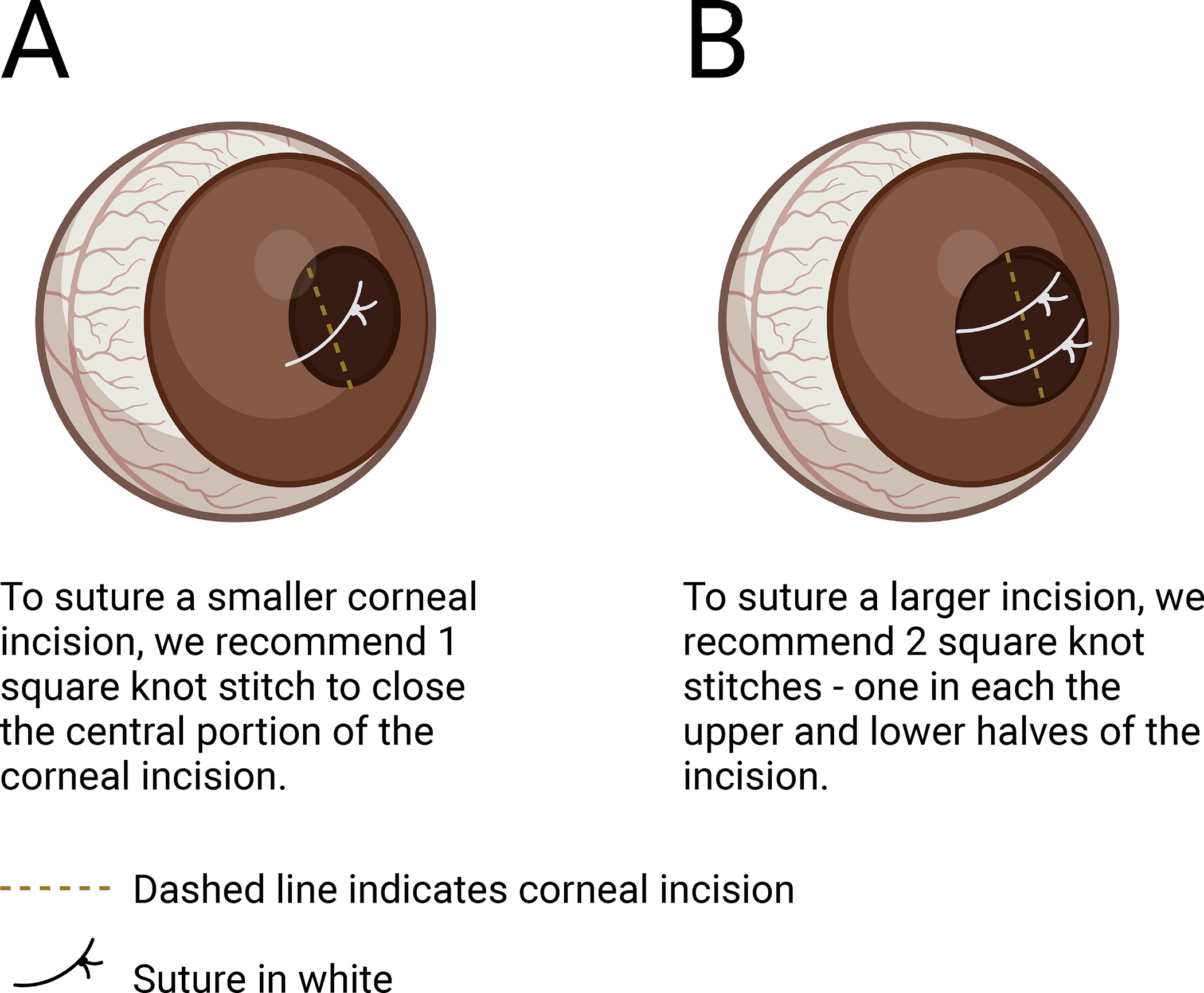

Figures

Figure 1:
Figure 1:. Schematic of the in vivo surgical technique that mimics cataract surgery in a mouse model.
Created with BioRender.com
Figure 2:
Figure 2:. Alternative approaches for extracting the lens fiber mass from the lens capsule.
The nuclear mass can be removed in 2 approaches either using the (A) needle/syringe apparatus or (B) micro forceps. Created with BioRender.com
Figure 3:
Figure 3:. Alternative approaches for suturing the corneal incision.
The number of stitches required to properly close the corneal opening depends on the size of the initial corneal incision. These approaches will close either (A) a small corneal incision using 1 square knot stitch or (B) a larger corneal incision using 2 square knot stitches, one square knot stitch in the upper and lower sections of the original incision. Created with BioRender.com
Figure 4:
Figure 4:. Visualization of the critical steps to model cataract surgery in mice.
Here we depict the following key steps of the protocol: (A) the typical size of a dilated mouse pupil when it is ready for surgical manipulation; (B) a corneal incision; (C) what the lens nucleus and fiber mass look like outside of the ocular cavity, as well as how to identify an intact capsular bag; (D,E) suturing of the corneal flaps; and lastly, (F) how the final square knot stitch should close the corneal gap.
Figure 5:
Figure 5:. Injury response time course in lens epithelial cells PCS in a mouse model.
This represents data collected from various published immunostaining and RNA sequencing experiments,,,,,. Staining was analyzed using embedded post-surgical and ‘time zero’ control mouse eyes. RNA sequencing was performed on mouse capsular bags post-surgery. Created with BioRender.com.
Figure 6:
Figure 6:. Efficacy of cataract surgery model.
(A) Volcano plot of genes differentially expressed in LECs at 6 h PCS. Blue dots represent negative fold change, red dots represent positive fold change, and yellow dot represents Il19. (B) Successful implementation of this technique has been used in studying the acute response of LECs following CS evident in this immunostaining of classic inflammatory markers like CXCL1 (red), S100a9 (red), and G-CSF (red), which upregulate as early as 3 h PCS with a more robust response observed at 6 h PCS; nuclei stained with Draq5 (blue). (C) Additionally, further efficacy of this surgical model is evident in this immunostaining of pSMAD3 (red) in LECs PCS, which correlates with F4/80-positive macrophage infiltration (green) in lens capsular bags. Detectable pSMAD3 is observed at 48 h PCS with peak pSMAD3-positive nuclei at 5 days PCS. Merge: pSMAD3 (red); F4/80 (green); nuclei stained with Draq5 (blue). Scale bar: 100 μm. Abbreviations: e, remnant lens epithelial cells/lens cells; lc, lens capsule. Panel A has been reprinted with permission from Novo et al.. Panel B and Panel C have been reprinted with permission from Jiang et al..

References

    1. Wormstone IM, Wormstone YM, Smith AJO, Eldred JA Posterior capsule opacification: What’s in the bag? Prog Retin Eye Res. 82, 100905 (2021). - PubMed
    1. Bassnett S, Shi Y, Vrensen GF Biological glass: structural determinants of eye lens transparency. Philos Trans R Soc Lond B Biol Sci. 366 (1568), 1250–1264 (2011). - PMC - PubMed
    1. Danysh BP, Duncan MK The lens capsule. Exp Eye Res. 88 (2), 151–164 (2009). - PMC - PubMed
    1. Liu YC, Wilkins M, Kim T, Malyugin B, Mehta JS Cataracts. Lancet. 390 (10094), 600–612 (2017). - PubMed
    1. Khairallah M et al. Number of people blind or visually impaired by cataract worldwide and in world regions, 1990 to 2010. Invest Ophthalmol Vis Sci. 56 (11), 6762–6769 (2015). - PubMed

Publication types

LinkOut - more resources