. 2023 Dec;6(6):502-513.
doi: 10.1089/crispr.2023.0039.

CRISPRi-Mediated Treatment of Dominant Rhodopsin-Associated Retinitis Pigmentosa

Affiliations

CRISPRi-Mediated Treatment of Dominant Rhodopsin-Associated Retinitis Pigmentosa

Erin R Burnight et al. CRISPR J. 2023 Dec.

Abstract

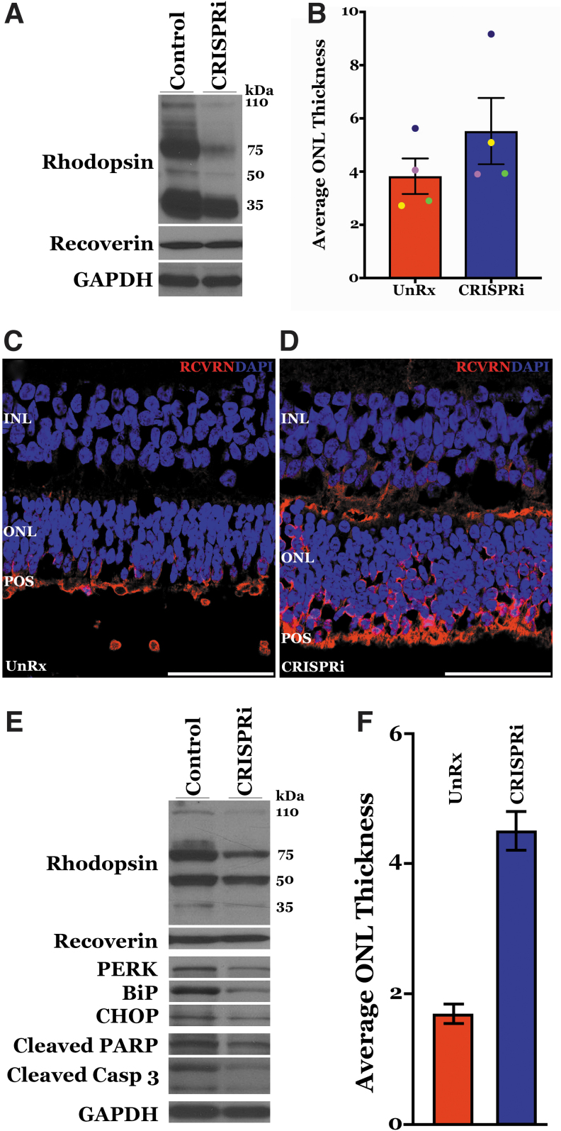
Rhodopsin (RHO) mutations such as Pro23His are the leading cause of dominantly inherited retinitis pigmentosa in North America. As with other dominant retinal dystrophies, these mutations lead to production of a toxic protein product, and treatment will require knockdown of the mutant allele. The purpose of this study was to develop a CRISPR-Cas9-mediated transcriptional repression strategy using catalytically inactive Staphylococcus aureus Cas9 (dCas9) fused to the Krüppel-associated box (KRAB) transcriptional repressor domain. Using a reporter construct carrying green fluorescent protein (GFP) cloned downstream of the RHO promoter fragment (nucleotides -1403 to +73), we demonstrate a ∼74-84% reduction in RHO promoter activity in RHOpCRISPRi-treated versus plasmid-only controls. After subretinal transduction of human retinal explants and transgenic Pro23His mutant pigs, significant knockdown of rhodopsin protein was achieved. Suppression of mutant transgene in vivo was associated with a reduction in endoplasmic reticulum (ER) stress and apoptosis markers and preservation of photoreceptor cell layer thickness.


Figures

FIG. 1.
FIG. 1.
CRISPRi-based transcriptional repression of RHO promoter. (A) Schematic of developed RHOp-CRISPRi plasmid used in these studies. (B) Diagram of the RHO promoter GFP reporter plasmid and Staphylococcus aureus guides targeting regulatory elements. Guides were designed using the Benchling platform and cloned into an AAV transgene cassette construct carrying S. aureus dCas9–KRAB. (C) Histogram showing GFP expression in HEK293T cells cotransfected with each guide and reporter construct relative to reporter transfection alone. GFP expression was determined using SYBR Green quantitative real-time PCR and standard curve analysis. Total number of GFP copies was normalized to number of 18S copies in each sample and knockdown was calculated as the percentage of GFP/18S ratios in controls treated with reporter plasmid alone. Significance was determined through two-tailed Student's t test. **p < 0.01; ****p < 0.0001. N = 3. AAV, adeno-associated virus; KRAB, Krüppel-associated box; PCR, polymerase chain reaction; RHO, rhodopsin.
FIG. 2.
FIG. 2.
CRISPRi machinery successfully targets and decreases rhodopsin protein expression in human retinal explants. (A) Reverse Transcriptase-PCR (RT-PCR) with (+) or without (−) reverse transcriptase detecting RHO and dCasKRAB transcript in each of three AAV5–RHOpCRISPRi-treated versus untreated human explants. Endogenous 18S transcript was detected as a control. (B) Western blot comparing whole tissue lysates from untreated (UnRx) versus AAV5–RHOpCRISPRi-treated (CRISPRi) human retinal explants ex vivo. Blots were probed using anti-RHO, anti-RCVRN, and anti-GAPDH primary antibodies. Anti-RHO produced three major species of rhodopsin antibody complexes at 35, 75, and 110 kDa corresponding to monomeric, dimeric, and trimeric forms of rhodopsin, respectively. (C) Semiquantitative densitometric analysis (RHO/GAPDH) between explant treatment groups. Colored dots correspond to untreated and AAV5–RHOpCRISPRi-treated explants cultured from the same human donor eye. Error bars represent the SEM and the asterisk (*) denotes statistical significance (p < 0.05). (D, E) Representative immunofluorescent micrographs comparing sections from untreated (D) and AAV5–RHOpCRISPRi-treated (E) explants labeled with anti-RHO (green). Cell nuclei are counterstained with DAPI (blue). Scale bar = 100 μm. AAV5, adeno-associated virus serotype 5; RCVRN, recoverin; SEM, standard error of the mean.
FIG. 3.
FIG. 3.
Genome-wide off-target transcript suppression analysis. (A) Number of differentially expressed transcripts between treatment versus control and donor 1 versus donor 2. (B, C) Volcano plots demonstrating that there are more significantly differentially expressed genes (shown in red) between tissue donors than between treatment and control conditions. Top differentially expressed genes are labeled. Enhanced volcano plots demonstrating that there is significantly more differences in gene expression between tissue donors than there is between treatment and control conditions.
FIG. 4.
FIG. 4.
Number of transgene copies and phenotypic analysis of the Pro23His rhodopsin transgenic pig model. (A) Digital PCR analysis of four P23H transgenic animals (2M and 2F—93-7) using probe-based assays targeted to human RHO. Number of copies was normalized to that of pig RNASEH1. Orange dotted line represents average of 5.6 ± 0.2 copies of human RHO. (B–D) Fluorescent confocal micrographs of retinas from wild-type (B) and P23H transgenic mini-swine (C, D) immunohistochemically labeled with anti-RHO (green) and stained with PNA (red) to label rod and cone photoreceptors, respectively. Scale bar = 50 μm. PNA, peanut agglutinin.
FIG. 5.
FIG. 5.
AAV5–RHOpCRISPRi subretinal delivery mitigates endoplasmic reticulum (ER) stress and preserves ONL thickness in Pro23His transgenic swine model. (A) Representative Western blot of retinal lysates from one animal demonstrating RHO repression in AAV5–RHOpCRISPRi vector-treated eye compared with untreated control eye 2 weeks post-transduction. (B) Histogram showing average number of nuclei per column in control and treated groups. Nuclei were counted using immunofluorescent confocal images of cryosectioned retinae from treated and untreated eyes (N = 4 animals). Error bars represent SEM. (C, D) Representative confocal images comparing sections from untreated (C) and AAV5–RHOpCRISPRi-treated (D) eyes at 2 weeks post-transduction labeled with anti-RCVRN (red). Cell nuclei are counterstained with DAPI (blue). (E) Western blot of retinal lysates from one animal demonstrating RHO repression in AAV5–RHOpCRISPRi vector-treated eye compared with untreated control eye 12 weeks posttransduction. ER stress markers PERK, BiP, CHOP, cleaved PARP, and cleaved Caspase 3 were reduced in lysate from the treated eye compared with that from the untreated contralateral eye. (F) Histogram showing average number of nuclei in control versus treated eye. Nuclei were counted using immunofluorescent confocal images of cryosectioned retinae from treated and untreated eyes (N = 4 animals). Error bars represent SEM. Scale bar = 50 μm. ONL, outer nuclear layer.

References

    1. Stone EM, Andorf JL, Whitmore SS, et al. . Clinically focused molecular investigation of 1000 consecutive families with inherited retinal disease. Ophthalmology 2017;124(9):1314–1331. - PMC - PubMed
    1. Hartong DT, McGee TL, Sandberg MA, et al. . Search for a correlation between telomere length and severity of retinitis pigmentosa due to the dominant rhodopsin Pro23His mutation. Mol Vis 2009;15:592–597. - PMC - PubMed
    1. Saliba RS, Munro PM, Luthert PJ, et al. . The cellular fate of mutant rhodopsin: Quality control, degradation and aggresome formation. J Cell Sci 2002;115(Pt 14):2907–2918. - PubMed
    1. Russell S, Bennett J, Wellman JA, et al. . Efficacy and safety of voretigene neparvovec (AAV2-hRPE65v2) in patients with RPE65-mediated inherited retinal dystrophy: A randomised, controlled, open-label, phase 3 trial. Lancet 2017;390(10097):849–860. - PMC - PubMed
    1. LaVail MM, Yasumura D, Matthes MT, et al. . Ribozyme rescue of photoreceptor cells in P23H transgenic rats: Long-term survival and late-stage therapy. Proc Natl Acad Sci U S A 2000;97(21):11488–11493. - PMC - PubMed