. 2023 Dec;113(6):331-351.
doi: 10.1007/s11103-023-01393-x. Epub 2023 Dec 18.

Cell wall anisotropy plays a key role in Zea mays stomatal complex movement: the possible role of the cell wall matrix

Affiliations

Cell wall anisotropy plays a key role in Zea mays stomatal complex movement: the possible role of the cell wall matrix

K Gkolemis et al. Plant Mol Biol. 2023 Dec.

Abstract

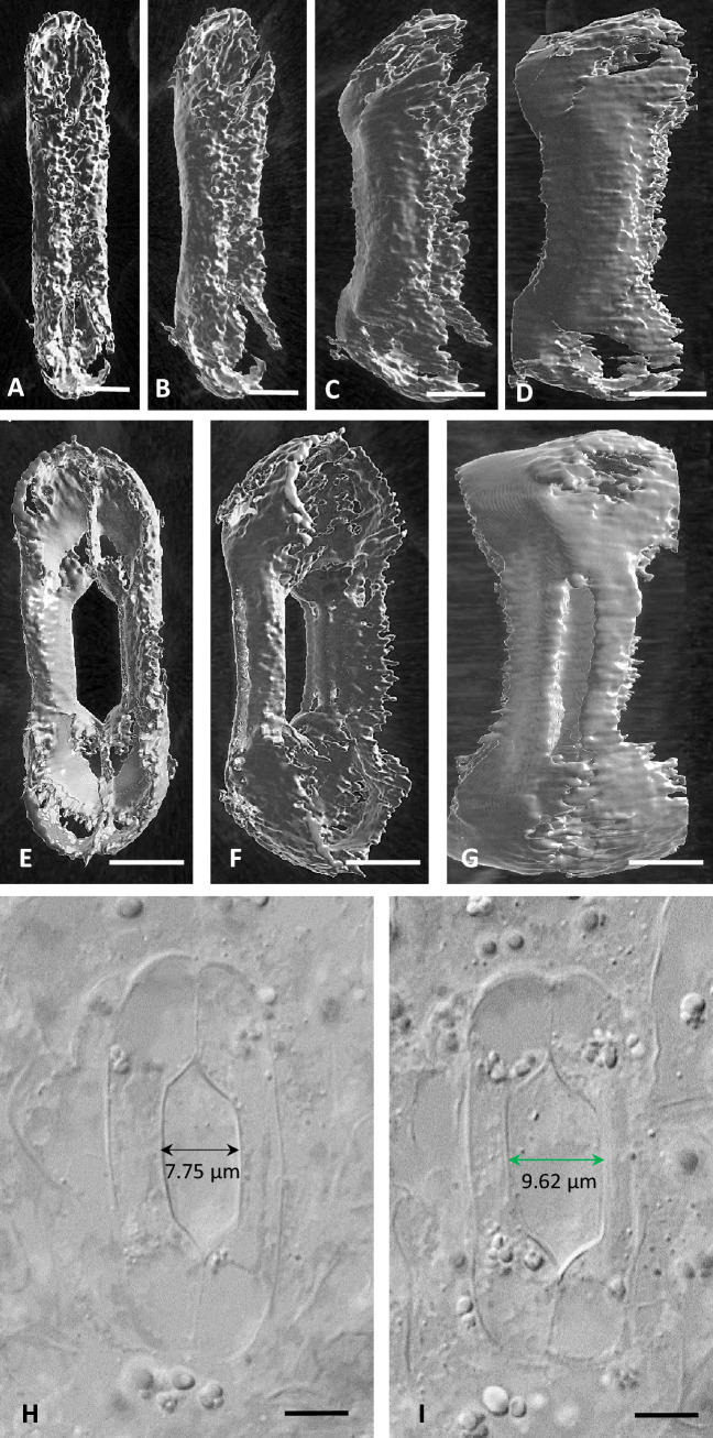
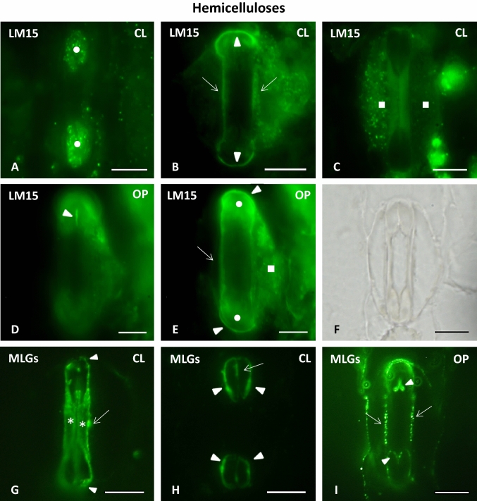
The opening of the stomatal pore in Zea mays is accomplished by the lateral displacement of the central canals of the dumbbell-shaped guard cells (GCs) towards their adjacent deflating subsidiary cells that retreat locally. During this process, the central canals swell, and their cell wall thickenings become thinner. The mechanical forces driving the outward displacement of the central canal are applied by the asymmetrically swollen bulbous ends of the GCs via the rigid terminal cell wall thickenings of the central canal and the polar ventral cell wall (VW) ends. During stomatal pore closure, the shrinking bulbous GC ends no longer exert the mechanical forces on the central canals, allowing them to be pushed back inwards, towards their initial position, by the now swelling subsidiary cells. During this process, the cell walls of the central canal thicken. Examination of immunolabeled specimens revealed that important cell wall matrix materials are differentially distributed across the walls of Z. mays stomatal complexes. The cell walls of the bulbous ends and of the central canal of the GCs, as well as the cell walls of the subsidiary cells were shown to be rich in methylesterified homogalacturonans (HGs) and hemicelluloses. Demethylesterified HGs were, in turn, mainly located at the terminal cell wall thickenings of the central canal, at the polar ends of the VW, at the lateral walls of the GCs and at the periclinal cell walls of the central canal. During stomatal function, a spatiotemporal change on the distribution of some of the cell wall matrix materials is observed. The participation of the above cell wall matrix polysaccharides in the well-orchestrated response of the cell wall during the reversible movements of the stomatal complexes is discussed.

Keywords: Anisotropy; Cell wall expansion; Cell wall matrix; Homogalacturonans; Stomatal complexes; Zea mays.


Conflict of interest statement

The authors declare no conflict of interest.

Figures

Fig. 1
Fig. 1
A Diagrammatic representation of a closed Z. mays stomatal complex in median paradermal view (from Galatis 1980). The dots represent microtubules. CC Central Canal, PVWE Polar Ventral Cell Wall End, LW Lateral Cell Wall, TW Transverse Cell Wall, SC Subsidiary Cell, VW Ventral Cell Wall. B (Ia) Diagrammatic representation of an external paradermal section of area I of Figure A. The arrowheads point to terminal cell wall thickenings of the central canal. (Ib) Diagrammatic representation of a transverse section of a stomatal complex. The section corresponds to the plane II of Figure A. The arrowheads point to the terminal cell wall thickenings of the central canal. (IIa) Diagrammatic representation of the stomatal complex in transverse section corresponding to the middle of the central canal (plane III in A). The intense cell wall thickenings of the central canal are shown. SC Subsidiary Cell. (IIb) Diagrammatic representation of an opening stomatal complex. The section corresponds to the middle of the central canal. Compare to IIa. SC Subsidiary Cell. C Paradermal section of Z. mays stomatal complex as depicted in TEM. The asterisks mark the bulbous ends of the GCs. The arrowheads point to the parts of the ventral cell wall between the central canal (CC) and the polar ends of the ventral cell wall (PVWE). SC subsidiary cell. Scale bar: 5 μm. D Transverse GC section as seen in TEM. The section corresponds to plane II of A. The asterisk mark the terminal cell wall thickenings of the central canal. Scale bar: 2 μm. E GC as depicted in TEM in transverse section passing through the middle of the central canal. Note the intense cell wall thickenings (asterisks). Scale bar: 2 μm. F Terminal cell wall thickening of the central canal as depicted in TEM in longitudinal section. Note the cellulose microfibrils orientation. Scale bar: 1 μm
Fig. 2
Fig. 2
A, B: Closed (A) and open (B) stomatal complex as seen in DIC in paradermal view. The arrowheads mark parts of the VW that elongate during the opening of the stomatal pore. SC Subsidiary Cell, SP Stomatal Pore. Scale bars: 10 μm. C, D: Transverse sections of a closed stomatal complex (C) and an opening stomatal complex (D) as seen in DIC. The sections pass through a median plane of the central canal. SC Subsidiary Cell. Scale bars: 5 μm. E Diagrammatic representation of a closed and an open stomatal complex. 1 width of bulbous end (BE width), 2 width of central canal (CC width), 3 width of subsidiary cell (SC width), 4 overall width of stomatal complex (StCo width), θ° angle between the ventral wall of the central canal and the ventral wall of the bulbous end, ω° angle between the lateral wall of the central canal and the lateral cell wall of the bulbous end. F Graphic comparison of the mean values of measured morphological parameters in closed (blue columns) and open stomata (red columns): i BE width and CC width, ii ratio of BE width to CC width, iii angles θ° and ω°, iv ratio of SC width to StCo width. Error bars on each column represent standard error. Numbers included in parentheses in the legends of the horizontal axis refer to each parameter’s numbering in E. Different letters state a statistically significant difference (P ≤ 0.001)
Fig. 3
Fig. 3
AD Successive captures of 3D reconstruction of a closed stoma using pictures taken by BC-43 confocal microscope. EG: Successive captures of 3D reconstruction of an open stoma using pictures taken by BC-43 confocal microscope. H, I: Paradermal sections of an opened Z. mays stoma corresponding to the middle of the GCs (H) and near the substomatal cavity (I), as seen in DIC optics. The width of the stomatal pore at these two planes is shown. Scale bars: 5 μm
Fig. 4
Fig. 4
Immunolabeling of highly methylesterified HG epitopes LM20-HG (A, B, E, F, I, J, K) and JIM7-HG (C, D, G, H, L) in paradermal sections of closed (AD) and open (EL) stomatal complexes. CL closed, OP open. Intense fluorescent signal is emitted by the periclinal cell walls of the central canal (asterisks in A), the lateral cell walls of the GCs (arrows in B, C, E, L), the transverse cell walls of the GCs (arrowheads in B, C, I, L) the polar VW ends (arrows in F, H), the periclinal cell walls of the subsidiary cells (squares in G, J) and the anticlinal cell walls of the subsidiary cells (arrows in D and arrowhead in K). Note that LM20-HG deposition is observed in the periclinal cell walls of the central canal of closed stomata (A), while the periclinal cell walls of the subsidiary cells are enriched with both LM20-HG (J) and JIM7-HG (G) epitopes in open stomata. Scale bars: 10 μm
Fig. 5
Fig. 5
Immunolabeling of demethylesterified HG epitopes JIM5-HG (A, B, E, F, I, J) and 2F4-HG (C, D, G, H, K, L) in paradermal sections of closed (AD, H) and open (EG, IL) stomatal complexes. CL closed, OP open. Intense fluorescent signal is emitted by the periclinal (asterisks in F and arrows in G) and the anticlinal (arrows in A, D, I, K) cell walls of the central canal, the terminal cell wall thickenings of the central canal (arrowheads in E, H), the transverse cell walls of the GCs (arrowheads in B, D, J), parts of the VW (arrowheads in G, L) and the periclinal (squares in C) and the anticlinal cell walls (arrows in B, J) of the subsidiary cells. Note that strong 2F4-HG immunofluorescence is exhibited by the periclinal cell walls of the subsidiary cells of closed stomata (C), while in open stomata the periclinal cell walls of the central canal are enriched in JIM5-HG (F) and 2F4-HG (G) alike. SP Stomatal Pore. Scale bars: 10 μm
Fig. 6
Fig. 6
HG epitopes in the periclinal cell walls of the central canal and of the subsidiary cells in closed and open stomata of Z. mays. AD: Diagrammatic representation of the distribution of pectin epitopes in the periclinal cell walls of the central canal and of the subsidiary cells in closed and open stomata. LM20 (A) is shown in red, JIM7 (B) in orange, JIM5 (C) in green and 2F4 (D) in blue. The cell wall thickenings of the central canal appear in gray. Signal detected at the terminal thickenings of the central canal is also depicted (C, D). E, F: Quantification of fluorescence expressed as corrected integrated density/μm2 in the periclinal cell walls of the central canal (E) and of the subsidiary cells (F) in closed and open stomata after incubation with antibodies LM20 (red), JIM7 (orange), JIM5 (green) and 2F4 (blue). Corrected integrated density/μm2 is measured in arbitrary fluorescence units (afu)/μm2 set by ImageJ
Fig. 7
Fig. 7
Immunolabeling of xyloglucans (LM15 epitope) (AE) and MLGs (BG-I) in paradermal sections of closed (AC, G, H) and open (D, E, I) stomatal complexes. CL closed, OP open. Intense LM15 fluorescent signal is emitted by the periclinal cell walls of the bulbous ends (circles in A, E), the polar VW ends (arrowheads in D and arrows in H), the lateral GC cell walls (arrows in B, E), the transverse GC cell walls (arrowheads in B, E) and the periclinal cell walls of the subsidiary cells (squares in C, E). BG-1 fluorescent signal is exhibited by the anticlinal (arrow in G), the lateral (arrow in I) and the transverse GC walls (arrowheads in G, signal also visible in H, I), the periclinal cell walls of the central canal (asterisks in G), the terminal cell wall thickenings of the central canal (arrowheads in H) and parts of the VW (arrows in H and arrowheads in I). F: the stomatal complex of I as seen in DIC optics. Scale bars: 10 μm
Fig. 8
Fig. 8
Closed (A, C, E) and open (B, D, F) stomatal complexes of three grasses species as seen in paradermal sections observed by DIC optics. Scale bars: 10 μm

References

    1. Apostolakos P, Giannoutsou E, Galatis B. Callose: a multifunctional (1, 3)-β-d-glucan involved in morphogenesis and function of angiosperm stomata. J Biol Res-Thessaloniki. 2021;28:17. doi: 10.1186/s40709-021-00150-9. - DOI - PMC - PubMed
    1. Aylor DE, Parlange J-Y, Krikorian AD. Stomatal mechanics. Am J Bot. 1973;60:163–171. doi: 10.1002/j.1537-2197.1973.tb10213.x. - DOI
    1. Bidhendi AJ, Geitmann A. Relating the mechanics of the primary plant cell wall to morphogenesis. J Exp Bot. 2016;67:449–461. doi: 10.1093/jxb/erv535. - DOI - PubMed
    1. Bidhendi AJ, Geitmann A. Finite element modeling of shape changes in plant cells. Plant Physiol. 2018;176:41–56. doi: 10.1104/pp.17.01684. - DOI - PMC - PubMed
    1. Buckeridge MS, Rayon C, Urbanowicz B, Tiné MAS, Carpita NC. Mixed linkage (13), (1 4)-d-glucans of grasses. Cereal Chem. 2004;81:115–127. doi: 10.1094/CCHEM.2004.81.1.115. - DOI

LinkOut - more resources