. 2023 Dec 18;33(3):e4870.
doi: 10.1002/pro.4870. Online ahead of print.

Exploring the conformational changes of the Munc18-1/syntaxin 1a complex

Affiliations

Exploring the conformational changes of the Munc18-1/syntaxin 1a complex

Ioanna Stefani et al. Protein Sci. .

Abstract

Neurotransmitters are released from synaptic vesicles, the membrane of which fuses with the plasma membrane upon calcium influx. This membrane fusion reaction is driven by the formation of a tight complex comprising the plasma membrane N-ethylmaleimide-sensitive factor attachment receptor (SNARE) proteins syntaxin-1a and SNAP-25 with the vesicle SNARE protein synaptobrevin. The neuronal protein Munc18-1 forms a stable complex with syntaxin-1a. Biochemically, syntaxin-1a cannot escape the tight grip of Munc18-1, so formation of the SNARE complex is inhibited. However, Munc18-1 is essential for the release of neurotransmitters in vivo. It has therefore been assumed that Munc18-1 makes the bound syntaxin-1a available for SNARE complex formation. Exactly how this occurs is still unclear, but it is assumed that structural rearrangements occur. Here, we used a series of mutations to specifically weaken the complex at different positions in order to induce these rearrangements biochemically. Our approach was guided through sequence and structural analysis and supported by molecular dynamics simulations. Subsequently, we created a homology model showing the complex in an altered conformation. This conformation presumably represents a more open arrangement of syntaxin-1a that permits the formation of a SNARE complex to be initiated while still bound to Munc18-1. In the future, research should investigate how this central reaction for neuronal communication is controlled by other proteins.

Keywords: Munc13; Munc18-1; SM protein; SNARE protein; neuronal secretion; synaptobrevin; syntaxin-1a.


Conflict of interest statement

The authors declare no competing interests.

Figures

FIGURE 1
FIGURE 1
Munc18‐1 forms a tight complex with syntaxin‐1a. (a) Cartoon representation of the structure of the Munc18‐1/syntaxin‐1a complex ((pdb:3c98; Burkhardt et al., ; Misura et al., 2000). (b) Domain organization of the two proteins, using the same color code as in the structure. The short N‐peptide (N) motif, the three Habc helices, the SNARE motif (H3), and the transmembrane region are indicated. Note that the H3 domain of syntaxin‐1a, which forms an extended helix in the SNARE complex (Sutton et al., 1998), has been further subdivided into Regions a, b, and c, in which bends are observed. The H3c region is buried within the central cavity of Munc18‐1 (Burkhardt et al., ; Misura et al., 2000).
FIGURE 2
FIGURE 2
The linker region of syntaxin‐1a is not essential for the interaction with Munc18‐1. (a) The linker region of syntaxin‐1a forms a short helix that interacts mostly with the H3‐ and Hc‐helices. Key residues mutated in the study are shown as sticks. On top, the conservation of the linker region is shown as a Weblogo representation (Crooks et al., 2004). Highly conserved residues are indicated. (b) Syntaxin‐1a linker mutants form a stable complex with Munc18‐1, as shown by native gel electrophoresis. Equimolar concentrations (100 μmol) of Munc18‐1 and syntaxin‐1a mutants were loaded individually or mixed as indicated. The band of the Munc18‐1/syntaxin‐1a complex is indicated by an arrow. Note that Munc18‐1 by itself does not form a defined band. (c) Addition of syntaxin‐1a to Munc18‐1 leads to an increase in the tryptophan‐emitted fluorescence. The emission spectra of Munc18‐1 alone, or in complex with Syxwt or SyxΔLinker, were recorded upon excitation at 295 nm. (d) Ternary SNARE complex formation for several syntaxin‐1a linker variants in the presence of M18wt, as observed by fluorescence anisotropy. Here, 40 nM of synaptobrevin labeled with Oregon Green at Cys28 were mixed with 500 nM syntaxin‐1a, preincubated with 750 nM Munc18‐1wt. SNARE complex formation was followed by an increase in fluorescence anisotropy upon the addition of 750 nM SNAP‐25. Fluorescence anisotropy was monitored at a wavelength of 524 nm upon excitation at 496 nm. When Syxwt (in gray) was preincubated with Munc18‐1, the SNARE complex formed slowly (black trace). Different Syntaxin‐1a linker mutants (SyxLE: light green, SyxΔlinker: green) were able to form a SNARE complex more rapidly than Syxwt in the presence of Munc18‐1. Note that we also mutated another highly conserved residue of the hydrophobic network of the linker region, F177 (SyxF177A). This single point mutation also somewhat released the inhibition of Munc18‐1 (Figure S2B). A double mutant, SyxR142A_L165A, which interfered with the polar and the hydrophobic networks on both side of the linker helix, was somewhat more disruptive than the individual exchanges (Figure S2C).
FIGURE 3
FIGURE 3
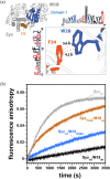
Munc18‐1 W28 interacts through pi–pi stacking with syntaxin‐1a F34. (a) In the Munc18‐1/syntaxin‐1a complex, the aromatic rings of syntaxin‐1a F34 and Munc18‐1 W28 lie within ~5 Å d and are oriented at a dihedral angle of ~30°. Both W28 and F34 are highly conserved across vertebrates, as shown by the Weblogo representations. (b) Mutations of both residues to alanines led to faster formation of the SNARE complex. Mixing experiments were carried out as described in the legend of Figure 2.
FIGURE 4
FIGURE 4
The N‐peptide is tethered to the Habc domain by a short stretch. (a) Weblogo representation of the short stretch that links the Habc domain to the N‐peptide of syntaxin 1a. Above, the section of the structure shows the position of this short region in the Munc18‐1/Syntaxin‐1a complex. The stretch is depicted as a dashed line, as its structure has not been resolved (Burkhardt et al., ; Misura et al., 2000). (b) In the presence of Munc18‐1, SyxΔΝ (orange yellow), Syx3x(11–26) (orange), and SyxΔ(11–26) (purple) formed a SNARE complex more rapidly than Syxwt (black). Mixing experiments were carried out as described in the legend of Figure 2. At first glance, the effect of the deletion of the short stretch between the N‐peptide and Habc domain (Syx∆11–26) may be surprising, but our subsequent analyses have shown that this short region also contributes to the interaction of syntaxin and Munc18‐1 (Table S3). The deletion could also have led to potentially undesirable contacts between the N‐peptide helix, which could not reach its normal binding site, and Domain 1 of Munc18‐1 or syntaxin1a itself, and change the dynamics of binding between Munc18 and syntaxin1a.
FIGURE 5
FIGURE 5
Munc18 α11α12 loop residues contribute to inhibition. (a) Different conformations of the α11α12 helical hairpin of Domain 3a, shown by an overlay of the Munc18‐1/syntaxin‐1a complex (pdb: 3c98, yellow), Munc18‐3 alone (pdb: 3puk, green), and Munc18‐1K332E_K333E (pdb: 6lpc, violet, top; Burkhardt et al., ; Hu et al., ; Misura et al., ; Wang et al., 2020). In the Munc18‐1/syntaxin‐1a complex, the region of Residues 317–333 is folded upwards towards the α11α12 helices. Note, that the stretch between Residues 317–323 was not resolved. In Munc18 alone, the α11α12 loop adopts an unfurled conformation with an extension of α12. The very conserved proline at the tip of α12 is thought to be the hinge point for the unfurling of the helix (bottom). The conservation of the Munc18‐1 helical hairpin loop is shown by a Weblogo representation. Mutated residues are indicated by arrows. (b) Complex formation of M18P335A (55 μmol) with Syxwt and different syntaxin‐1a variants (34 μmol) in the absence and presence of the SNARE partners SNAP‐25 and synaptobrevin (each 90 μmol), monitored by native gel electrophoresis. The positions of the monomeric proteins and complexes are indicated by arrows. Note that synaptobrevin cannot be detected in the nondenaturing gel because of its isoelectric point. Although a clear complex band was visible for the M18P335A/Syxwt complex, the complexes of M18P335A/Syxwt complex appeared as more diffuse bands, suggesting that these interactions were less stable. (c) Deletion of the hairpin loop, Munc18‐1Δ317–333, had only a very small effect on Munc18‐1's ability to inhibit the formation of the SNARE complex, as monitored by fluorescence anisotropy. In contrast, the mutation of the conserved P335 to an alanine rendered Munc18‐1 much less able to control the complex formation of the bound syntaxin‐1a, in agreement with previous studies (Han et al., ; Munch et al., ; Parisotto et al., ; Park et al., ; Wang et al., 2020). Mixing experiments were carried out as described in the legend of Figure 2. (d) Determination of the off‐rate of the Munc18‐1/syntaxin‐1a complex by competitive dissociation. An excess of unlabeled Syx1a variants (5 μM) was added to a premix of 100 nM Oregon Green‐labeled Syx1a variants using the single cysteine introduced at Position 1 along with 250 nM Munc18‐1; and the decrease in fluorescence anisotropy. was measured. The dissociation was fitted by a single exponential. Dissociation was faster for the Domain 3a variants (M18P335A, M18Δ(317–333), M18Y337A) than for M18wt. The dissociation rates are given in Table S1.
FIGURE 6
FIGURE 6
Model of Munc18‐1/Syntaxin‐1a in an intermediate conformation. (a) Cartoon representation of the Munc18‐1/syntaxin‐1a complex modeled by using the Vps45/Tlg2 structure as a template. (b) Overlay of the modeled structure of the Munc18‐1/syntaxin‐1a complex in a more open conformation (color code as in a) and the crystal structure (in white).

Similar articles

References

    1. Abraham MJ, Murtola T, Schulz R, Páll S, Smith JC, Hess B, et al. GROMACS: high performance molecular simulations through multi‐level parallelism from laptops to supercomputers. SoftwareX. 2015;1–2:19–25.
    1. Abramov D, Guiberson NGL, Burré J. STXBP1 encephalopathies: clinical spectrum, disease mechanisms, and therapeutic strategies. J Neurochem. 2021;157:165–178. - PMC - PubMed
    1. Anderson RJ, Weng Z, Campbell RK, Jiang X. Main‐chain conformational tendencies of amino acids. Proteins. 2005;60:679–689. - PubMed
    1. André T, Classen J, Brenner P, Betts MJ, Dörr B, Kreye S, et al. The interaction of Munc18‐1 helix 11 and 12 with the central region of the VAMP2 SNARE motif is essential for SNARE Templating and synaptic transmission. eNeuro. 2020;7:ENEURO.0278‐ENEU20.2020. - PMC - PubMed
    1. Archbold JK, Whitten AE, Hu S‐H, Collins BM, Martin JL. SNARE‐ing the structures of Sec1/Munc18 proteins. Curr Opin Struct Biol. 2014;29:44–51. - PubMed

LinkOut - more resources