. 2024 Jan;43(1):3-14.
doi: 10.14366/usg.23116. Epub 2023 Aug 29.

Artificial intelligence in breast ultrasound: application in clinical practice

Affiliations

Artificial intelligence in breast ultrasound: application in clinical practice

Hila Fruchtman Brot et al. Ultrasonography. 2024 Jan.

Abstract

Ultrasound (US) is a widely accessible and extensively used tool for breast imaging. It is commonly used as an additional screening tool, especially for women with dense breast tissue. Advances in artificial intelligence (AI) have led to the development of various AI systems that assist radiologists in identifying and diagnosing breast lesions using US. This article provides an overview of the background and supporting evidence for the use of AI in hand held breast US. It discusses the impact of AI on clinical workflow, covering breast cancer detection, diagnosis, prediction of molecular subtypes, evaluation of axillary lymph node status, and response to neoadjuvant chemotherapy. Additionally, the article highlights the potential significance of AI in breast US for low and middle income countries.

Keywords: Artificial intelligence; Breast neoplasms; Computer-aided detection; Computer-aided diagnosis; Ultrasound.


Conflict of interest statement

No potential conflict of interest relevant to this article was reported.

Figures
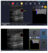
Fig. 1.
Fig. 1.. BR-FHUS Navigation - an adjunct artificial intelligence tool to assist breast ulrasoundscreening.
A. The software provides a route map panel to display scanning information including probe location and scanning coverage (scanned areas of the breast indicated by gray bars). In addition, the system generates a "lesion detection module" to assist with real-time detection. Example of a suspicious lesion (red square) detected by the software with display of clock axis and distance to the nipple (purple circle) is shown. The images and cine loops are stored with their spatial position coordinates in DICOM format (figure provided by TaiHao Medical Inc. and used with permission). B. BR-FHUS Viewer assists physicians in reviewing a series of 2-D ultrasound images recorded by BR-FHUS Navigation. It supports the computer-aided detection method to detect breast lesions in the recorded images. An example of a snapshot taken by Navigation which is automatically showed in the Viewer so physicians can choose which lesion would appear in the report. By clicking a CADe button (not displayed on this image) the software automatically detects the suspicious areas which are displayed in the suspicious area list (purple rectangle), indicating breast laterality (green rectangle) and location within the breast (yellow rectangle) for each lesion. The user can capture the image which suits for reporting (in this case marked by blue background from the list area on the right side). Subsequently, the diagnostics results including DICOM images and reports are uploaded to PACS or stored in a local storage (figure provided by TaiHao Medical Inc. and used with permission). BR-FHUS, breast free-hand ultrasound; CADe, computer assisted detection; PACs, Picture Archiving and Communication System.
Fig. 2.
Fig. 2.. Example of BU-CAD showing orthogonal B-mode ultrasound images of a breast mass (indicated by the red box), where the artificial intelligence decision support output (DS) displayed a high score of lesion characteristics (SLC=61), corresponding to a Breast Imaging Reporting and Data System (BI-RADS) assessment category 4B (figure provided by TaiHao Medical Inc. and used with permission).
Fig. 3.
Fig. 3.. Example of Koios decision support (DS) for Breast showing the B-mode ultrasound images of invasive carcinoma with micropapillary features in a 68-year-old patient.
The artificial intelligence DS output is displayed in a graphical form on the right panel, with the DS-generated output (in this case correctly classified as "suspicious") and the confidence of assessment within that category as marked by the triangular marker.
Fig. 4.
Fig. 4.. Example of S-Detect showing the B-Mode ultrasound image of breast mass.
The artificial intelligence decision support output outlined the region of interest around the mass margins (yellow line); classified descriptors as irregular shape, non-parallel orientation, spiculated margin, and heterogenous echo pattern. S-Detect produced a final assessment of "possibly malignant” supporting a recommendation to biopsy this finding (figure provided by Samsung Electronics Co., Ltd.).

References

    1. Boyd NF, Guo H, Martin LJ, Sun L, Stone J, Fishell E, et al. Mammographic density and the risk and detection of breast cancer. N Engl J Med. 2007;356:227–236. - PubMed
    1. Wanders JO, Holland K, Veldhuis WB, Mann RM, Pijnappel RM, Peeters PH, et al. Volumetric breast density affects performance of digital screening mammography. Breast Cancer Res Treat. 2017;162:95–103. - PMC - PubMed
    1. Lee JM, Arao RF, Sprague BL, Kerlikowske K, Lehman CD, Smith RA, et al. Performance of screening ultrasonography as an adjunct to screening mammography in women across the spectrum of breast cancer risk. JAMA Intern Med. 2019;179:658–667. - PMC - PubMed
    1. Berg WA, Blume JD, Cormack JB, Mendelson EB, Lehrer D, Bohm-Velez M, et al. Combined screening with ultrasound and mammography vs mammography alone in women at elevated risk of breast cancer. JAMA. 2008;299:2151–2163. - PMC - PubMed
    1. Mendelson EB, Bohm-Velez M, Berg WA, Whitman GJ, Feldman MI, Madjar H, et al. In: ACR BI-RADS Atlas, Breast Imaging Reporting and Data System. D'Orsi CJ, Sickles EA, Mendelson EB, Morris EA, editors. Reston, VA: American College of Radiology; 2013. ACR BI-RADS ultrasound; pp. 1–154.

LinkOut - more resources