. 2024 Feb;626(7998):435-442.
doi: 10.1038/s41586-023-06953-1. Epub 2023 Dec 18.

De novo design of high-affinity binders of bioactive helical peptides

Affiliations

De novo design of high-affinity binders of bioactive helical peptides

Susana Vázquez Torres et al. Nature. 2024 Feb.

Abstract

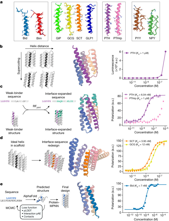
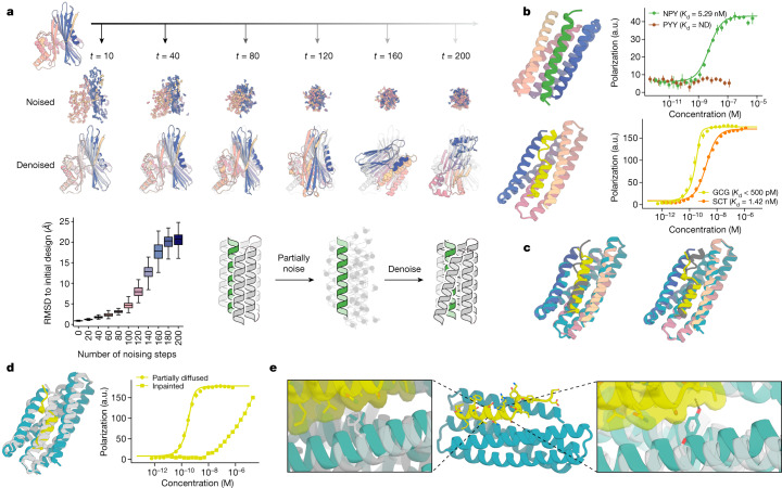
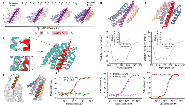
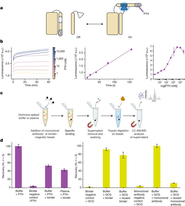
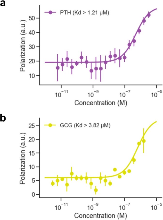
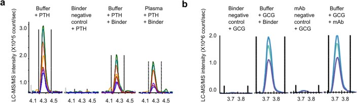
Many peptide hormones form an α-helix on binding their receptors1-4, and sensitive methods for their detection could contribute to better clinical management of disease5. De novo protein design can now generate binders with high affinity and specificity to structured proteins6,7. However, the design of interactions between proteins and short peptides with helical propensity is an unmet challenge. Here we describe parametric generation and deep learning-based methods for designing proteins to address this challenge. We show that by extending RFdiffusion8 to enable binder design to flexible targets, and to refining input structure models by successive noising and denoising (partial diffusion), picomolar-affinity binders can be generated to helical peptide targets by either refining designs generated with other methods, or completely de novo starting from random noise distributions without any subsequent experimental optimization. The RFdiffusion designs enable the enrichment and subsequent detection of parathyroid hormone and glucagon by mass spectrometry, and the construction of bioluminescence-based protein biosensors. The ability to design binders to conformationally variable targets, and to optimize by partial diffusion both natural and designed proteins, should be broadly useful.


Conflict of interest statement

D.B., S.V.T., P.J.Y.L., P.V., I.D.L., A.N.H., D.J., E.H., A.H.-W.Y., H.-H.H., J.L.W., M.J.M., N.R.B. and G.R.L. are inventors on a provisional patent application submitted by the University of Washington for the design and composition of the proteins created in this study.

Figures

Fig. 1
Fig. 1. Design strategies for binding helical peptides.
a, Helical peptide targets: apoptosis-related BH3 domains of Bid (PDB ID: 4QVE) and Bim (PDB ID: 3FDL), GCG (PDB ID: 1GCN), gastric inhibitory peptide (GIP; PDB ID: 2QKH), SCT (PDB ID: 6WZG), GCG-like peptide 1 (GLP1; PDB ID: 6X18), PTH (PDB ID: 1ET1), PTHrp (PDB ID: 7VVJ), PYY (PDB ID: 2DEZ) and NPY (PDB ID: 7X9A). b, Parametric approach. Left: sampling groove scaffolds varying supercoiling and helix distance to fit different targets. Middle: design model (spectrum) and PTH target (purple) of the best parametrically designed PTH binder. Right: split NanoBiT titration of PTH and the binder showed weak binding. a.u., arbitrary units. c, Inpainting binder optimization. Left: redesign of parametrically generated binder designs using RFjoint Inpainting to expand the binding interface and ProteinMPNN to redesign the sequences. Middle: AF2 prediction of Inpainted design (spectrum) with extended interface (teal), and PTH target (purple). Right: FP measurements (n = 4) indicate 6.04 nM binding to PTH and weak binding to off-target PTHrp. d, Threading approach to peptide binder design. Left: starting with a helix-bound scaffold, a target is threaded onto the bound helix and the interface is redesigned. Middle: AF2 prediction of design (spectrum) and SCT target (orange). Right: FP measurements (n = 4) indicate 3.95 nM binding to SCT and 12 nM binding to GCG. e, Hallucinating peptide binders. Left: Markov chain Monte Carlo (MCMC) steps are carried out in sequence space. At each step, the peptide sequence is re-predicted, and changes are accepted or rejected on the basis of interfacial contacts and AF2 metrics. The final structure is then redesigned using ProteinMPNN to avoid adversarial sequences. Interaction pAE, predicted alignment error across the interface; pLDDT, predicted local distance difference test. Middle: AF2 prediction of design (spectrum) and Bid target (blue). Right: FP measurements (n = 4) indicate 7 nM binding affinity to Bid.
Fig. 2
Fig. 2. Peptide binder optimization with RFdiffusion.
a, Top: partial diffusion. RFdiffusion is used to denoise a randomly noised starting design (left); varying the extent of initial noising (middle row) enables control over the extent of introduced structural variation (bottom row; colours, new designs; grey, original design). Bottom left: partial diffusion diversifies designs. Note that the greater the amount of noise added, the more dissimilar the outputs are to the starting structure. Bottom right: depiction of the helix binder optimization strategy. b, Top: design model (spectrum) of the partially diffused binder to NPY (green) and FP measurements (n = 4) indicating a 5.3 nM binding affinity to NPY target and selectivity over PYY (brown). ND, not detectable. Bottom: design model (spectrum) of the partially diffused binder to GCG (yellow) and FP measurements (n = 4) indicating a subnanomolar binding affinity to GCG and selectivity over SCT (orange). c, Left: model (spectrum with GCG in grey) aligns with 0.72 Å RMSD to the 1.95-Å crystal structure (teal and yellow) of the RFjoint Inpainted GCG binder. Right: model (spectrum with GCG in grey) aligns with 0.6 Å RMSD to the 1.81-Å crystal structure (teal and yellow) of the partially diffused GCG binder. d, Left: the crystal structures of the Inpainted (grey) and partially diffused (teal and yellow) GCG binders have considerable topological similarity; there are many small readjustments. Right: FP titrations (n = 4) with GCG indicate much tighter binding following partial diffusion. e, Left inset: the crystal structure of the partially diffused backbone (teal) shows how the newly introduced Ile13 increases shape complementarity compared to the phenylalanine in the Inpainted binder (crystal structure in grey; structures aligned on residues 16–29 of GCG). Middle: crystal structure of the partially diffused GCG binder (teal and yellow). Right inset: the backbone shifts in the partially diffused structure (teal) enable Tyr16 to make packing and hydrogen-bonding interactions with the peptide; Ser16 in the original design did not make any peptide contacts (grey).
Fig. 3
Fig. 3. De novo peptide binder design with RFdiffusion.
a, Schematic showing peptide binder design using RFdiffusion. Starting from a random distribution of residues around the target peptide (XT), successive RFdiffusion denoising steps progressively remove the noise leading at the end of the trajectory to a folded structure, X0, cradling the peptide. At each step t, RFdiffusion predicts the final structure pX0 given the current noise sample Xt, and a step that interpolates in this direction is taken to generate the input for the next denoising step Xt−1. b, Design of picomolar-affinity PTH binder. Top: design model of PTH binder (spectrum, AF2 metrics in Supplementary Table 9). Middle: circular dichroism data show that the binder has helical secondary structure and is stable at 95 °C (inset). Bottom: FP measurements (n = 4) with PTH indicate a subnanomolar binding affinity and no binding to PTHrp indicates high specificity. c, Design of picomolar-affinity Bim binder. Top: design model of Bim binder (spectrum, AF2 metrics in Supplementary Table 9). Middle: circular dichroism data show that the binder has helical secondary structure and is stable at 95 °C (inset). Bottom: FP measurements (n = 4) with Bim indicate a subnanomolar binding affinity. d, Crystal structure of Bim binder (teal and red). Top inset: a cross-interface hydrogen-bond network formed between Asn20 of Bim and Thr73 and Asn77 of the binder. Bottom inset: a kinked helix in the diffused backbone accommodates Arg13 of Bim. e, RFdiffusion with PYY sequence input alone. Left: PYY in complex with its native NPY Y2 receptor (PDB ID: 7YON) shows flexibility at its N and C termini (teal). Middle: design model of the binder (spectrum) with PYY target (brown); the peptide is more ordered in both regions (N terminus, teal). Right: FP measurements (n = 4) with PYY indicate a 24.5 nM binding affinity.
Fig. 4
Fig. 4. Application of designed binders to sensing and detection.
a, The PTH lucCage biosensor. Cage and latch (left, beige), key (right, beige) and the PTH binder (grey) thermodynamically shift from the off to on state in the presence of PTH peptide target (purple). This conformational change brings two luciferase halves (inactive in white, active in blue) close together leading to luminescence. b, Left: titration of PTH results in luminescence increase (n = 3). Middle: response of lucCagePTH biosensor in the linear concentration range, indicating a 10 nM limit of detection (Supplementary Methods). Right: titration curve of 10 nM lucCagePTH + lucKey to various concentrations of PTH (n = 3). c, LC–MS/MS enrichment experiment schematic; the trypsin digestion step was skipped for the GCG binder. d, Left: LC–MS/MS recovery percentages for triplicate measurements of an N-terminal tryptic peptide of PTH. The negative control comprised bovine serum albumin mixed with PTH in a buffer solution. Right: recovery percentage for triplicate measurements of intact GCG peptide normalized to the percentage recovery with a monoclonal antibody (n = 3). Following the first binding and elution experiments, beads were extensively washed and resuspended in PBS–CHAPS 0.1%, and then used in a second pulldown experiment. An unrelated binder attached to the magnetic beads mixed with GCG in buffer was used as a negative control. a,c, Created with BioRender.com.
Extended Data Fig. 1
Extended Data Fig. 1. Low affinity RFjoint-Inpainted binders for NPY and GCG using extended parametric designs.
(a) Left: Design model (colour spectrum + yellow) of the tightest GCG binder. Right: FP titration (n = 4) for the tightest GCG binder indicates ~ 231 nM binding affinity (b) Left: Design model (colour spectrum + dark green) of the tightest NPY binder. Right: FP titration (n = 4) for the tightest NPY binder indicates 3.5 µM binding affinity.
Extended Data Fig. 2
Extended Data Fig. 2. Additional binders made using threading and redesign.
(a) Left: Design model (colour spectrum + dark blue) of the tightest GLP1 binder. Right: FP titration (n = 4) for the tightest GLP1 binder indicates 68.8 nM binding affinity (b) Left: Design model (colour spectrum + green) of the tightest GIP binder. Right: FP titration (n = 4) for the tightest GIP binder indicates 6.96 nM binding affinity.
Extended Data Fig. 3
Extended Data Fig. 3. Hallucinated Bid binders are stable and bind Bid peptide with high affinity.
(a) 46 Hallucinated designs tested for initial experimental screening. (b) 4 designs were chosen for expression without Bid peptide. All expressed as monomeric proteins (assessed by preparative SEC) and were pure by SDS-PAGE (n = 1). (c) All Hallucinations could be pulled-down by biotinylated Bid immobilised on streptavidin magnetic beads. B = bound to bead, U = unbound, in supernatant. L = ladder (n = 1). (d) Bid is unstructured in isolation by circular dichroism (CD), whereas all Hallucinations were helical in isolation, as predicted from the Hallucinated structure. A 1:1 molar ratio of binder:Bid (Mix) produced greater helical signal than that predicted by the isolated spectra (No inter.) suggesting binding is inducing helix formation (n = 1). (e) Melting with CD showed that Hallucinations were thermostable, and binding to Bid increased thermostability (where measurable) (n = 1). All Hallucinated binders would remain folded, or refold after heating and cooling, in contrast to the natural binder Mcl-1 which precipitated in the process. (f) ITC showed that Hallucinations bound to Bid, with µM to nM Kds (n = 1). (g) FP measurements of designed Bid binders (n = 3).
Extended Data Fig. 4
Extended Data Fig. 4. Partial diffusion increases designability of native proteins.
500 native proteins of length 100 to 300 residues were selected from the PDB (< 3.5 Å resolution and no missing residues). Three different methods were applied to these proteins: 1) single sequence AlphaFold2 (AF2), 2) ProteinMPNN combined with AF2, and 3) partial diffusion (60 steps, noise = 1), ProteinMPNN and AF2. (a) Partial diffusion generates diverse protein conformations from the initial fold while maintaining the same overall fold, as indicated by the TM (Template Modeling) score exceeding 0.5. (b) The backbones resulting from partial diffusion exhibit higher designability compared to the native backbone, implying that they have been idealised for design purposes. (c) Visualisation of an example where partial diffusion + ProteinMPNN results in a significantly more designable protein relative to sequence redesign by ProteinMPNN on the native backbone.
Extended Data Fig. 5
Extended Data Fig. 5. PTH and GCG binders designed with RFdiffusion.
Representative binding data is shown for PTH (a) and GCG (b) binders designed by providing sequence input alone. The binding affinities, as measured by FP (n = 4), indicate low micromolar interactions with the respective peptide targets.
Extended Data Fig. 6
Extended Data Fig. 6. LC-MS/MS chromatograms for PTH and GCG binders.
(a) LC-MS/MS chromatograms for SVSEIQLMHNLGK, the N-terminal tryptic peptide of PTH; different peptide fragments detected by the LC-MS/MS assay are in different colours. (b) LC-MS/MS chromatograms for the intact GCG peptide HSQGTFTSDYSKYLDSRRAQDFVQWLMNT; different peptide fragments detected by the LC-MS/MS assay are in different colours.

References

    1. Pioszak AA, Xu HE. Molecular recognition of parathyroid hormone by its G protein-coupled receptor. Proc. Natl Acad. Sci. USA. 2008;105:5034–5039. doi: 10.1073/pnas.0801027105. - DOI - PMC - PubMed
    1. Park C, et al. Structural basis of neuropeptide Y signaling through Y1 receptor. Nat. Commun. 2022;13:853. doi: 10.1038/s41467-022-28510-6. - DOI - PMC - PubMed
    1. Sasaki K, Dockerill S, Adamiak DA, Tickle IJ, Blundell T. X-ray analysis of glucagon and its relationship to receptor binding. Nature. 1975;257:751–757. doi: 10.1038/257751a0. - DOI - PubMed
    1. Fukuhara S, et al. Structure of the human secretin receptor coupled to an engineered heterotrimeric G protein. Biochem. Biophys. Res. Commun. 2020;533:861–866. doi: 10.1016/j.bbrc.2020.08.042. - DOI - PubMed
    1. Wewer Albrechtsen NJ, Kuhre RE, Pedersen J, Knop FK, Holst JJ. The biology of glucagon and the consequences of hyperglucagonemia. Biomark. Med. 2016;10:1141–1151. doi: 10.2217/bmm-2016-0090. - DOI - PubMed